Alimentary System
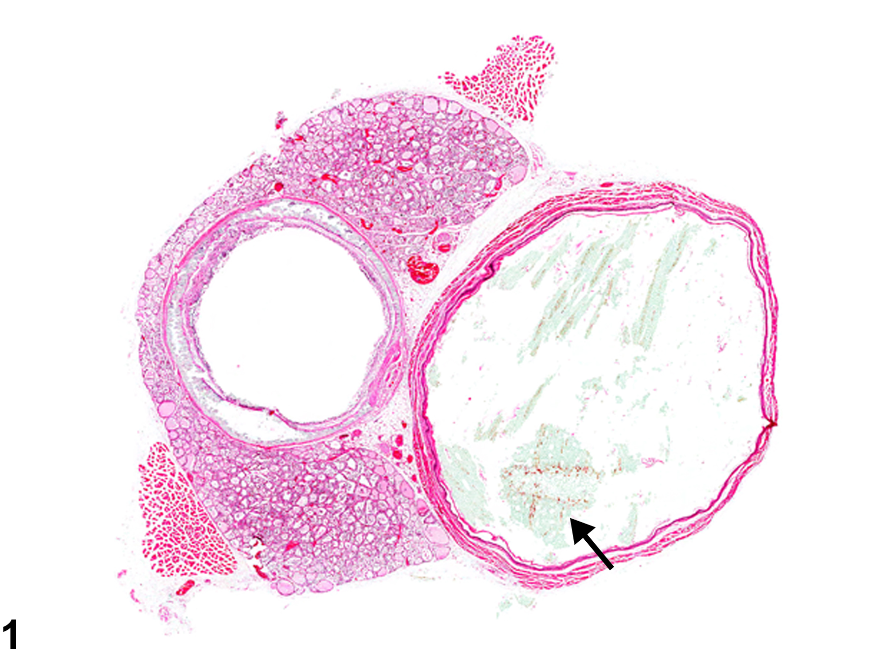
Esophagus - Dilation
Narrative
Brown HR, Hardisty JF. 1990. Oral cavity, esophagus and stomach. In: Pathology of the Fischer Rat (Boorman GA, Montgomery CA, MacKenzie WF, eds). Academic Press, San Diego, CA, 9-30.
Abstract: https://www.ncbi.nlm.nih.gov/nlmcatalog/9002563Harkness JE, Ferguson FG. 1979. Idiopathic megaesophagus in rat. Lab Anim Sci 49:495-498.
Abstract: https://www.ncbi.nlm.nih.gov/pubmed/513620
Esophagus - Dilation in a male Wistar Han rat from a chronic study. The dilated esophagus contains feed material (arrow).


