Reproductive System, Female
Oviduct - Necrosis
Narrative
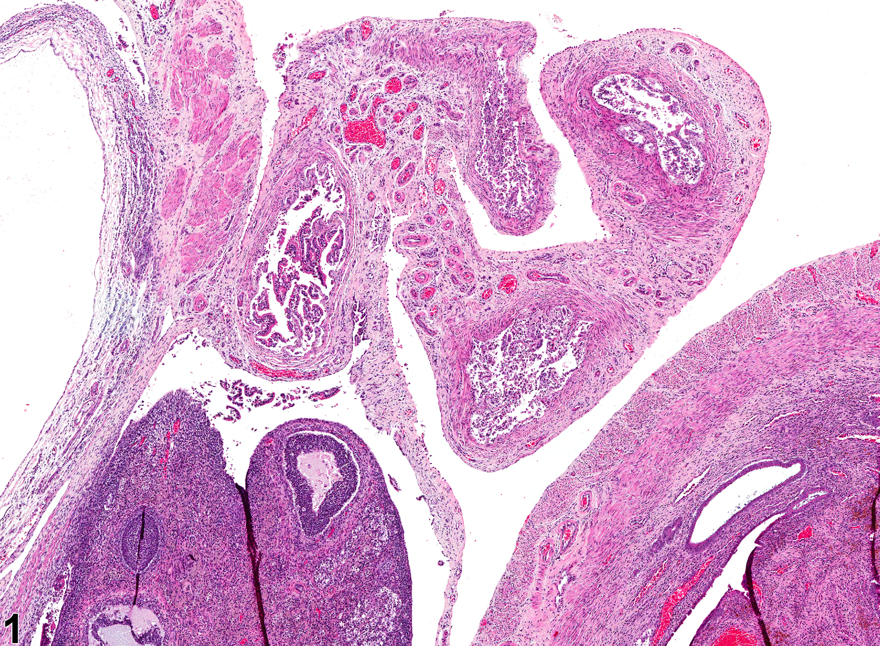
Oviduct necrosis is rarely seen in rodent studies. It may be a result of an associated inflammatory change in the adjacent ovary or uterus. Oviduct necrosis is characterized by hypereosinophilic, dissociated, fragmented, and sloughed epithelial cells (Figure 1 and Figure 2). Nuclei are pyknotic and karyorrhectic. It is usually accompanied by an inflammatory infiltrate.
Oviduct - Necrosis should be diagnosed and graded whenever present. Secondary lesions such as inflammation or hemorrhage should not be diagnosed unless warranted by severity. If the necrosis is secondary to another lesion, such as inflammation, it should not be diagnosed unless warranted by severity.
National Toxicology Program. 2006. NTP TR-521. Toxicology and Carcinogenesis Studies of 2,3,7,8-Tetrachlorodibenzo-p-dioxin (TCDD) (CAS No. 1746-01-6) in Female Harlan Sprague-Dawley Rats (Gavage Studies). NTP, Research Triangle Park, NC.
Abstract: https://ntp.niehs.nih.gov/go/9303National Toxicology Program. 2006. NTP TR-526. Toxicology and Carcinogenesis Studies of a Mixture of 2,3,7,8-Tetrachlorodibenzo-p-dioxin (TCDD) (CAS No. 1746-01-6), 2,3,4,7,8-Pentachlorodibenzofuran (PeCDF) (CAS No. 57117-31-4), and 3,3',4,4',5-Pentachlorobiphenyl (PCB 126) (CAS No. 57465-28-8) in Female Harlan Sprague-Dawley Rats (Gavage Studies). NTP, Research Triangle Park, NC.
Abstract: https://ntp.niehs.nih.gov/go/9307
Oviduct - Necrosis in a female Harlan Sprague-Dawley rat from a chronic study. There is sloughing of necrotic epithelial cells into the lumen of the oviduct.


