Reproductive System, Female
Clitoral Gland - Hyperplasia
Narrative
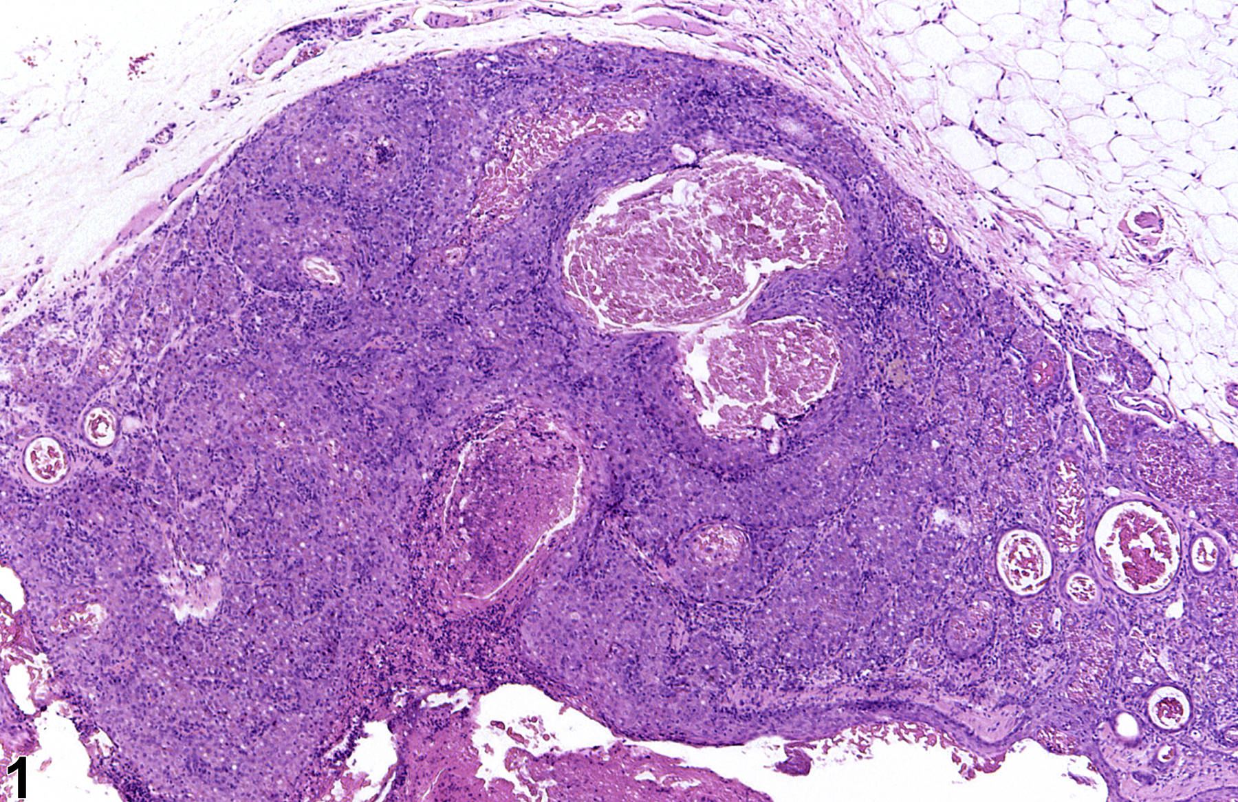
Clitoral gland hyperplasia is typically a focal lesion and more commonly occurs in the glandular tissue but may also occur in the ducts. The lesion may be unilateral or bilateral. Glandular hyperplasia is characterized by an increased number of acinar cells and may be accompanied by an increased number of basal cells. The acini are enlarged and appear crowded compared with the adjacent glandular tissue; however, there is normal cellular morphology and orientation. These latter features differentiate hyperplasia from adenomas, in which acinar structure is distorted, with frequent acinar fusion and solid clusters of neoplastic cells. Clitoral gland hyperplasia occurs as a spontaneous lesion and is often secondary to inflammation, especially in older animals. Hyperplasia is not considered a preneoplastic change in these older animals but, rather, a reactive hyperplasia, and there is no evidence of progression (note inflammation in Figure 1 and Figure 2). Androgenic drugs can induce epithelial hyperplasia of the clitoral gland.
Clitoral gland - Hyperplasia should be diagnosed whenever present and given a severity grade. Secondary lesions such as inflammation occurring within a focus of hyperplasia should not be diagnosed separately unless warranted by severity.
Copeland-Haines D, Eustis SL. 1990. Specialized sebaceous glands. In: Pathology of the Fischer Rat: Reference and Atlas (Boorman GA, Eustis SL, Elwell MR, Montgomery CA, MacKenzie WF, eds). Academic Press, San Diego, CA, 279-293.
Seely JC, Boorman GA. 1991. Mammary gland and specialized sebaceous glands. In: Pathology of the Mouse: Reference and Atlas (Maronpot RR, Boorman GA, Gaul BW, eds). Cache River Press, Vienna, IL, 613-635.
Stoll RE, Holden HE, Barthel CH, Blanchard KT. 1999. Oxymetholone: III. Evaluation in the p53+/- transgenic mouse model. Toxicol Pathol 27:513-518.
Abstract: https://www.ncbi.nlm.nih.gov/pubmed/10528630Stoll RE, Holden HE, Blanchard KT. 1999. Oxymetholone: II. Evaluation in the Tg-AC transgenic mouse model for detection of carcinogens. Toxicol Pathol 27:507-512.
Abstract: https://www.ncbi.nlm.nih.gov/pubmed/10528629
Clitoral gland - Hyperplasia in a female F344/N rat from a chronic study. An area of acinar hyperplasia is sharply demarcated from the adjacent clitoral gland; dilated ducts are also evident.


