Integumentary System
Skin - Fibrosis
Narrative
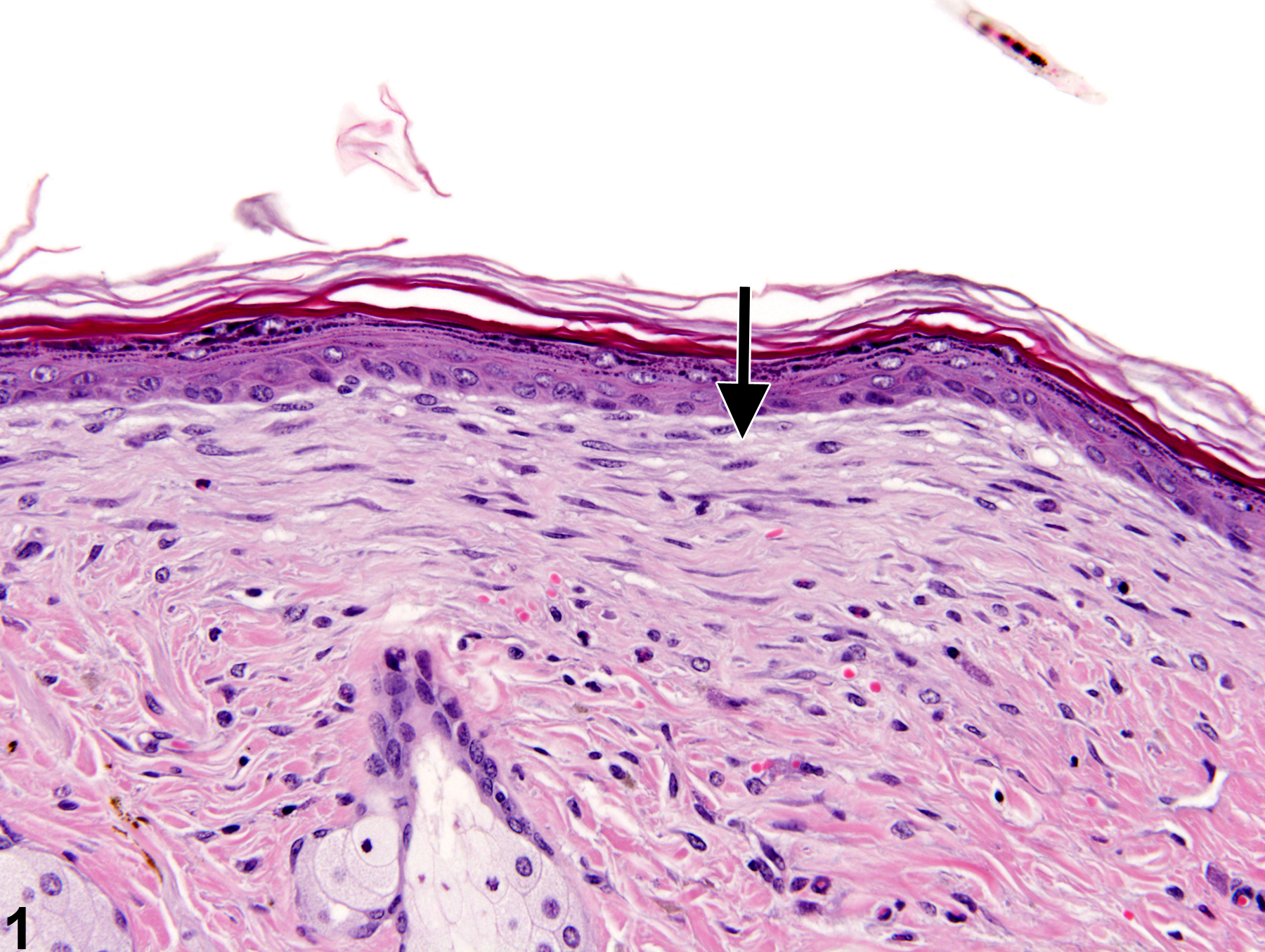
Fibrosis is often a sequela of epidermal or dermal injury due to chronic chemical exposure or trauma. It is characterized by an increase of fibrous connective tissues in the dermis (Figure 1) or subcutis and usually accompanies chronic inflammation. Fibrosis may occur as a subtle finding, particularly in the later stages. It is characterized by proliferation of fibroblasts and collagen fibers in the dermis or around hair follicles, typically oriented parallel to the epidermis. In more severe cases, the fibrosis can extend deeper into the dermis and subcutis. Areas of fibrosis may have a slightly basophilic appearance compared with the nonfibrotic dermis, but this can vary with staining quality and the maturity of the fibrotic lesion. In early fibrosis, the fibroblasts are usually accompanied by inflammatory cells (particularly when associated with an epidermal ulcer); the fibroblasts tend to be larger and more active, and the collagen fibers are less compact and more disorganized. In the later stages of fibrosis, the fibroblasts tend to be smaller and more spindle shaped; the collagen fibers are more organized and compact, and there is often less inflammation. In severe cases, there may be a loss of hair follicles in the fibrotic region.
Klein-Szanto AJP, Conti CJ. 2002. Skin and oral mucosa. In: Handbook of Toxicologic Pathology, 2nd ed (Haschek WM, Rousseaux CG, Wallig MA, eds). Academic Press, San Diego, 2:85-116.
Abstract: http://www.sciencedirect.com/science/book/9780123302151Peckham JC, Heider K. 1999. Skin and subcutis. In: Pathology of the Mouse: Reference and Atlas (Maronpot RR, Boorman GA, Gaul BW, eds). Cache River Press, Vienna, IL, 555-612.
Abstract: http://www.cacheriverpress.com/books/pathmouse.htm
Fibrosis-increase of fibrous connective tissues in the dermis (arrow) in a male B6C3F1 mouse from a 2-year study.


