Respiratory System
Nose, Epithelium - Regeneration
Narrative
Comment:
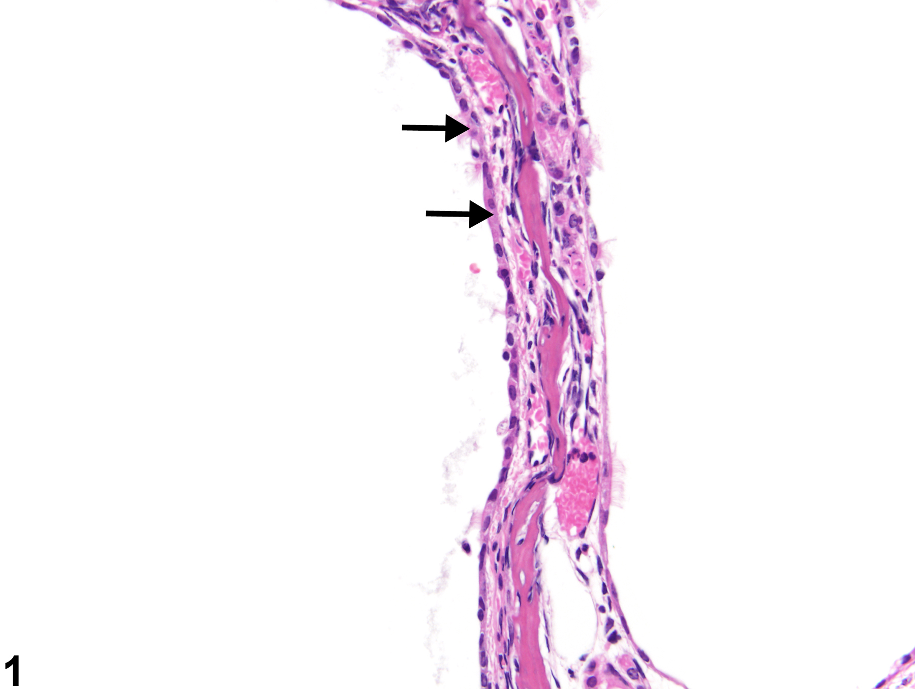
Regeneration (Figure 1) is the re-epithelialization of nasal structures following loss of the epithelium. The new epithelial layer is usually a thin layer of flattened, elongated cells that replaces the lost epithelium. The nuclei of these cells are often anisokaryotic, and there may seem to be fewer cells than there should be for the length of the defect. The cells are anisocytotic and lack normal polarity. The cytoplasm of involved cells is generally basophilic. Because it is a response to the loss of cells, regeneration is typically seen in areas of degeneration and/or necrosis, and it may coincide with hyperplasia and metaplasia.
Recommendations:
Regeneration should be diagnosed whenever present and evidence of necrosis or degeneration is also present. Regeneration should not be graded. To convey the severity of the lesion, either necrosis or degeneration must be diagnosed concurrently and graded (see
and
). The location of the lesion in the nasal cavity should be indicated in the diagnosis by including as a site modifier the type of epithelium that should be present (e.g., respiratory, olfactory). If the number of epithelial cells exceeds that which would be considered within normal limits, hyperplasia should be diagnosed, regardless of whether it is a direct effect of test article exposure or excessive regeneration, but the pathogenesis of the hyperplasia should be discussed in the pathology narrative. Other associated lesions, such as inflammation, atrophy, or metaplasia, should be diagnosed separately. If the regenerating epithelium exhibits significant features of atypia, based on the pathologist’s judgment, then "atypia, cellular" should be diagnosed and graded.
References:
Herbert RA, Leninger JR. 1999. Nose, larynx, and trachea. In: Pathology of the Mouse: Reference and Atlas (Maronpot RR, ed). Cache River Press, Vienna, IL, 259-289.
 Nose, Respiratory epithelium - Regeneration in a female B6C3F1/N mouse from a chronic study. A thin layer of epithelium is covering an area where the respiratory epithelium was previously lost (arrows).


