Reproductive System, Female
Vagina - Inflammation
Narrative
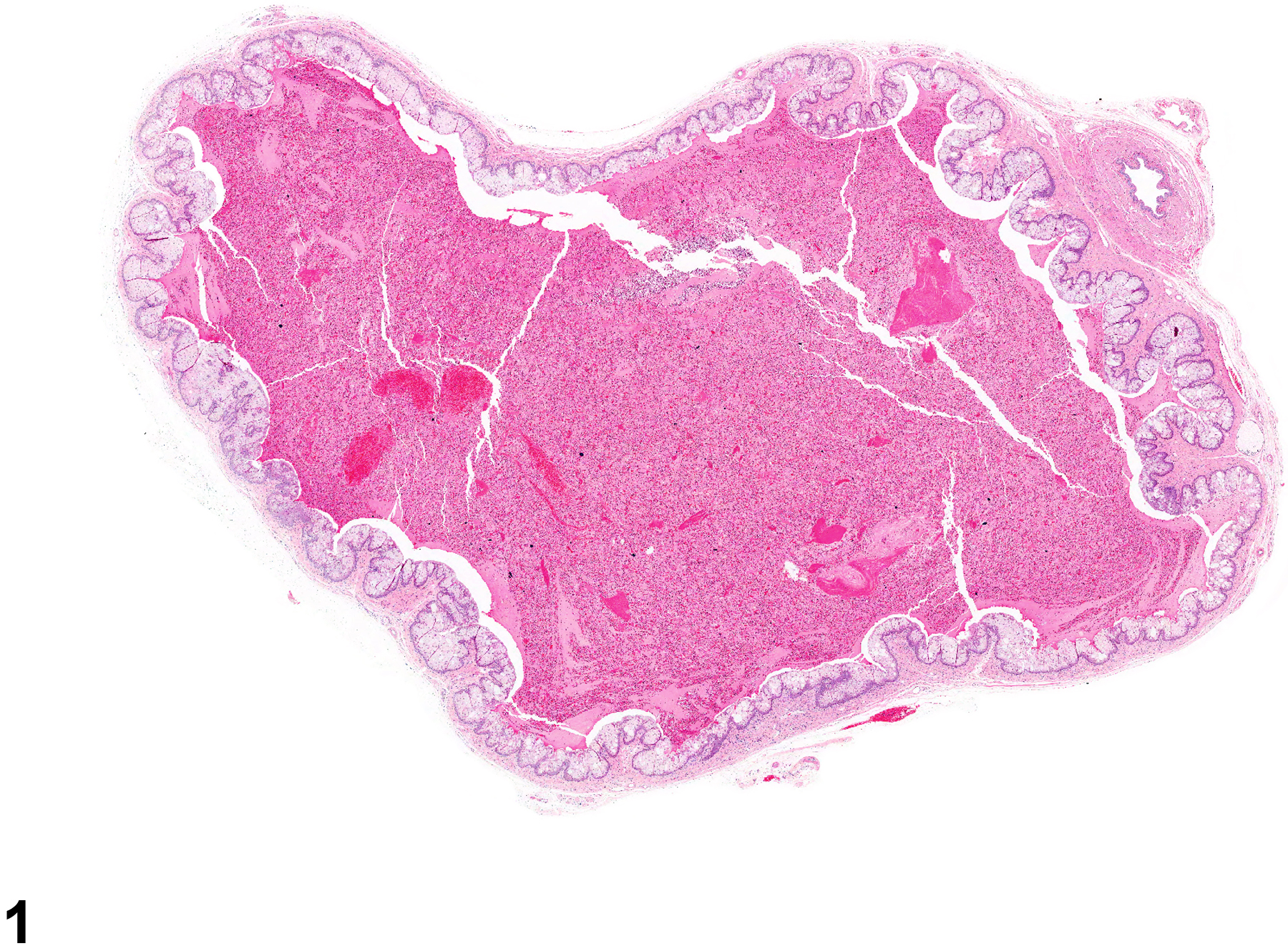
In NTP studies, there are five standard categories of inflammation: acute, suppurative, chronic, chronic active, and granulomatous. In acute inflammation (Figure 1 and Figure 2), the predominant infiltrating cell is the neutrophil, though fewer macrophages and lymphocytes may also be present. There may also be evidence of edema or hyperemia. The neutrophil is also the predominant infiltrating cell type in suppurative inflammation (Figure 3 and Figure 4), but they are aggregated, and many of them are degenerate (suppurative exudate). Cell debris, from both the resident cell populations and infiltrating leukocytes, and proteinaceous fluid containing fibrin, fewer macrophages, occasional lymphocytes or plasma cells, and, possibly, an infectious agent may also be present within the exudate. Grossly, these lesions would be characterized by the presence of pus. In the tissue surrounding the exudate, there may be fibroblasts, fibrous connective tissue, and mixed inflammatory cells, depending on the chronicity of the lesion. Abscesses should be diagnosed histologically as suppurative inflammation. Lymphocytes predominate in chronic inflammation. Lymphocytes also predominate in chronic active inflammation (Figure 5 and Figure 6), but there are also a significant number of neutrophils. Both lesions may contain macrophages. Granulomatous inflammation is another form of chronic inflammation, but this diagnosis requires the presence of a significant number of aggregated, large, activated macrophages, epithelioid macrophages, or multinucleated giant cells. Inflammation is differentiated from cellular infiltrates by the presence of other changes, such as edema, hemorrhage, degeneration, necrosis, or other evidence of tissue damage.
Frank suppurative inflammation (pyometra) is occasionally encountered in the vagina and is often associated with inflammation in the contiguous organs, cervix, and uterus. Mild inflammation is commonly observed, but this must be differentiated from the normal presence of inflammatory cells at certain stages in the estrous cycle. In the estrus cycle there tend to be more transepithelial migratory neutrophils, with only a few, if any, luminal accumulations of neutrophils.
Greaves P, Faccini JM. 1984. Female genital tract. In: Rat Histopathology: A Glossary for Use in Toxicity and Carcinogenicity Studies. Elsevier, Amsterdam, 171-179.
Leininger JR, Jokinen MP. 1990. Oviduct, uterus and vagina. In: Pathology of the Fischer Rat (Boorman GA, Eustis SL, Elwell MR, Montgomery CA, MacKenzie WF, eds). Academic Press, San Diego, CA, 443-459.
National Toxicology Program. 1989. NTP TR-342. Toxicology and Carcinogenesis Studies of Dichlorvos (CAS No. 62-73-7) in F344/N Rats and B6C3F1 Mice (Gavage Studies). NTP, Research Triangle Park, NC.
Abstract: https://ntp.niehs.nih.gov/go/10790National Toxicology Program. 2010. NTP TR-558. Toxicology and Carcinogenesis Studies of 3,3',4,4'-Tetrachloroazobenzene (TCAB) (CAS No. 14047-09-7) in Harlan Sprague-Dawley Rats and B6C3F1 Mice (Gavage Studies). NTP, Research Triangle Park, NC.
Abstract: https://ntp.niehs.nih.gov/go/33564Westwood FR. 2008. The female rat reproductive cycle: A practical histological guide to staging. Toxicol Pathol 36:375-384.
Abstract: https://www.ncbi.nlm.nih.gov/pubmed/18441260
Vagina - Inflammation, Acute in a female F344/N rat from a chronic study. The lumen of the vagina is filled with copious eosinophilic material.


