Reproductive System, Female
Ovary - Thrombosis
Narrative
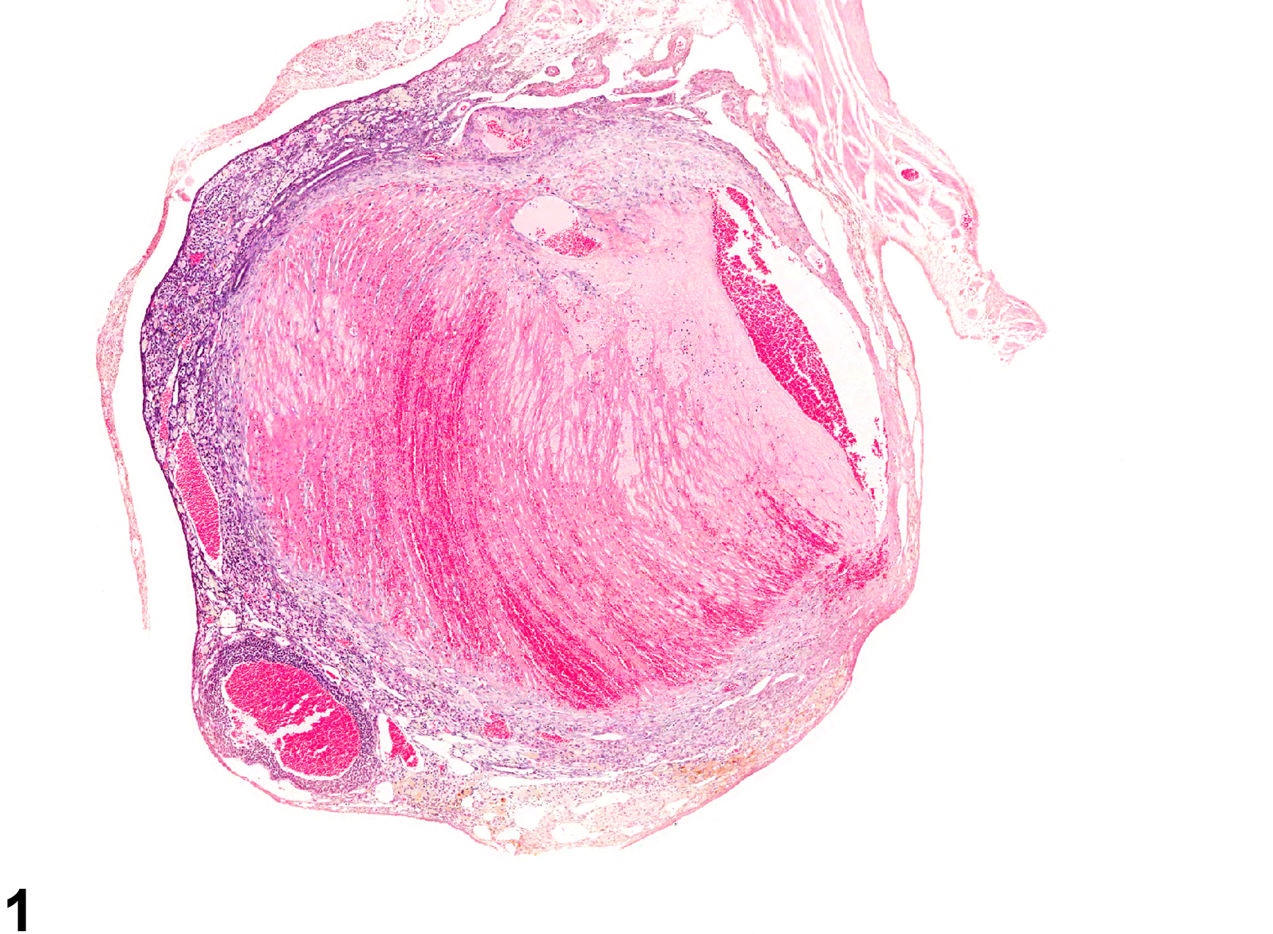
Ovarian thrombi (Figure 1 and Figure 2) may be large and comprise well-circumscribed areas of ovarian parenchyma that have been replaced by eosinophilic laminated material. The apparent laminations (lines of Zahn) are produced by alternating layers of platelets admixed with darker-staining fibrin, separated by darker layers containing mainly erythrocytes. Peripherally, at the attachment of the thrombus to the vessel wall, there are infiltrating fibroblasts and small numbers of inflammatory cells. There is compression of the adjacent parenchyma. Thrombi of longer duration are characterized by having less prominent lines of Zahn and often have clumps of amorphous basophilic material consistent with mineralization.
Three factors predispose to thrombosis: injury to endothelium, alterations in normal blood flow, and alterations in blood hypercoagulability. Endothelial injury may be due to trauma, infections, radiation, immune-mediated injuries, and bacterial toxins or endotoxins. Alterations in blood flow may occur in vessels narrowed by processes such as inflammation, atherosclerosis, medial mineralization, or medial hypertrophy that contribute to stasis or turbulence, which brings platelets into contact with the endothelium, and/or slow the inflow of clotting factor inhibitors. Alterations in blood coagulability (i.e., hypercoagulability) are not a common cause of thrombosis. However, they can be due to genetic defects in a coagulation protein or could be secondary to systemic disease (e.g., lymphoma, inflammation, renal disease).
Ovary - Thrombosis should be diagnosed but need not be graded. Necrosis secondary to thrombosis should be diagnosed and graded separately. Other lesions considered to be secondary to thrombosis (e.g., mineralization) should not be diagnosed separately unless warranted by severity.
Alison RH, Morgan KT, Montgomery CA. 1990. Ovary. In: Pathology of the Fischer Rat: Reference and Atlas (Boorman GA, Eustis SL, Elwell MR, Montgomery CA, MacKenzie WF, eds). Academic Press, San Diego, CA, 429-442.
Greaves P. 2012. Cardiovascular system. In: Histopathology of Preclinical Toxicity Studies: Interpretation and Relevance in Drug Safety Evaluation, 4th ed. Academic Press, San Diego, CA, 263-324.
Greaves P. 2012. Female genital tract. In: Histopathology of Preclinical Toxicity Studies: Interpretation and Relevance in Drug Safety Evaluation, 4th ed. Elsevier, Amsterdam, 667-724.
Maekawa A, Maita K, Harleman JH. 1996. Changes in the ovary. In: Pathobiology of the Aging Mouse (Mohr U, Dungworth DL, Capen CC, Carlton WW, Sundberg JP, Ward JM, eds). ILSI Press, Washington, DC, 451-467.
Mitchell RN. 2005. Hemodynamic disorders, thromboembolic disease, and shock. In: Robbins and
Cotran Basic Pathology of Disease, 7th ed (Vinay K, Abbas AK, Fausto N, eds). Elsevier Saunders, Philadelphia, PA, 119-144.
Montgomery CA, Alison RH. 1987. Non-neoplastic lesions of the ovary in Fischer 344 rats and B6C3F1 mice. Environ Health Perspect 73:53-75.
Abstract: https://www.ncbi.nlm.nih.gov/pmc/articles/PMC1474552/National Toxicology Program. 1993. NTP TR-404. Toxicology and Carcinogenesis Studies of 5,5-Diphenylhydantoin (CAS No. 57-41-0) (Phenytoin) in F344/N Rats and B6C3F1 Mice (Feed Studies). NTP, Research Triangle Park, NC.
Abstract: https://ntp.niehs.nih.gov/go/7688
Ovary - Thrombosis in a female B6C3F1/N mouse from a chronic study. A large thrombus occupies the majority of an ovary.


