Respiratory System
Nose - Foreign Material
Narrative
Comment:
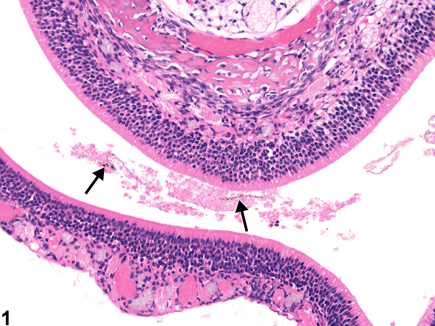
Foreign material (Figure 1, Figure 2, Figure 3, Figure 4, and Figure 5) refers to inhaled particulate test material or presumed test material. Therefore, this term applies only to inhalation studies or gavage studies in which the test material has been inhaled or exhaled into the nasal cavity. The term "pigmentation" is reserved for endogenous pigments (hemosiderin, lipofuscin) found in the nose and should not be used for exogenous material (see
). If the material has the expected morphologic appearance, is found only in treated animals, and increases in amount with increasing exposure level, it may be assumed to be the test agent. The morphologic appearance of the foreign material can be compared to the material found in the lung (Figure 5), where, in inhalation studies, it will usually be more abundant. The term “foreign body” is reserved for inhaled bits of plant material (feed or bedding), hair fragments, or other substances that are found in control and treated animals or do not increase in severity with increasing exposure level (see
).
It is uncommon to see the test agent in the nose. There may be little response to small amounts of inert particles, but more reactive materials may elicit an inflammatory response and changes in the nasal epithelium (e.g., degeneration or necrosis, metaplasia, atrophy).
It is uncommon to see the test agent in the nose. There may be little response to small amounts of inert particles, but more reactive materials may elicit an inflammatory response and changes in the nasal epithelium (e.g., degeneration or necrosis, metaplasia, atrophy).
Recommendations:
Foreign material (test material or presumed test material) should be diagnosed and graded whenever present. The specific location (e.g., respiratory or olfactory epithelium, nasopharyngeal duct) should be indicated in the diagnosis as a site modifier. If the foreign material is present in the nasal lumen or in more than one location, the site modifier may be omitted and the locations described in the pathology narrative. Associated lesions (e.g., inflammation, necrosis, fibrosis, epithelial hyperplasia) should be diagnosed and graded separately.
References:
References not listed.

Nose - Foreign material in a female F344/NTac rat from an acute study. Free dark brown particulate material is present in the nasal cavity (arrows).


