Reproductive System, Male
Preputial Gland - Atrophy
Narrative
Comment:
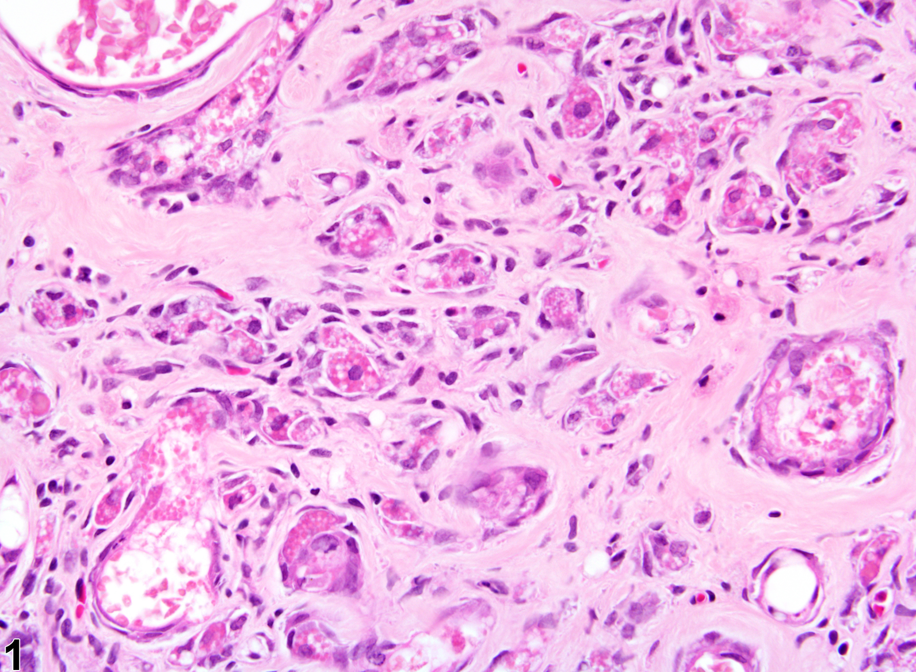
Preputial gland atrophy consists of reduction in the size of acini and reduction in the number of acinar epithelial cells comprising the atrophic acini. Prominent fibrosis may be associated with the acinar atrophy (Figure 1). Preputial gland atrophy may be a background lesion. It is often associated with chronic inflammation or other age-associated lesions.
Recommendations:
Preputial gland atrophy should be diagnosed and graded when it is a predominant change. If it is considered secondary to inflammation or another lesion, it should not be diagnosed separately unless warranted by severity. Likewise, associated lesions, if considered secondary to atrophy, should not be diagnosed separately unless warranted by severity. If atrophy is present in both glands, it should be qualified as bilateral.
References:
Boorman GA, Elwell MR, Mitsumori K. 1990. Male accessory sex glands, penis, and scrotum. In: Pathology of the Fischer Rat: Reference and Atlas (Boorman GA, Eustis SL, Elwell MR, Montgomery CA, MacKenzie WF, eds). Academic Press, San Diego, 419-428.
Abstract: http://www.ncbi.nlm.nih.gov/nlmcatalog/9002563