Reproductive System, Female
Vagina - Necrosis
Narrative
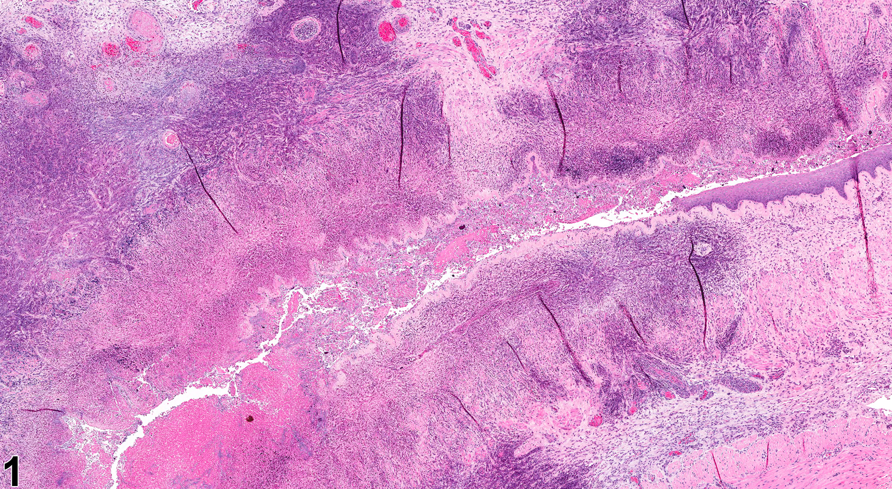
Necrosis of the vaginal epithelium (Figure 1 and Figure 2) is rarely observed. It is most typically observed secondary to intravaginal administration of xenobiotics, irritating vehicles, and/or intravaginal devices. The features of vaginal necrosis include nuclear pyknosis and/or karyorrhexis, cytoplasmic eosinophilia, cellular swelling or shrinkage, exfoliated cells, and/or intraluminal cellular debris. Vaginal necrosis is often associated with acute inflammation and may result in erosion/ulcer.
Necrosis of the vaginal epithelium and muscularis should be diagnosed and graded. If the necrosis is secondary to another lesion, such as inflammation or neoplasia, it should not be diagnosed separately. Lesions that are secondary to necrosis, such as inflammation, hemorrhage, or edema, should not be diagnosed separately unless warranted by severity but should be described in the pathology narrative as a component of the necrosis.
National Toxicology Program. 1994. NTP TR-433. Toxicology and Carcinogenesis Studies of Tricresyl Phosphate (CAS No. 1330-78-5) in F344/N Rats and B6C3F1 Mice (Gavage and Feed Studies). NTP, Research Triangle Park, NC.
Abstract: https://ntp.niehs.nih.gov/go/6010
Vagina - Necrosis, Epithelial in a female Harlan Sprague-Dawley rat from a chronic study. There is necrosis of the vagina.


