Reproductive System, Female
Vagina - Mucification
Narrative
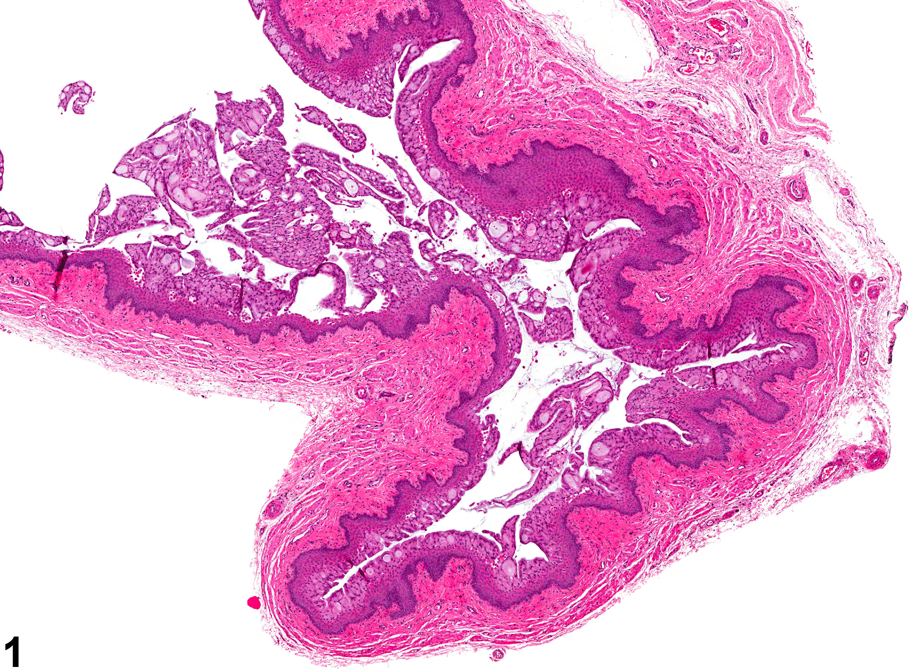
In general, vaginal mucification (Figure 1 and Figure 2) is induced by progesterone excess. Therefore, it occurs normally during proestrus, pregnancy, and pseudopregnancy. It is also seen with hyperprolactinemia, such as that caused by a prolactin-secreting pituitary neoplasm. It is characterized histologically by changes in the superficial epithelial cells of the vaginal mucosa, which become cuboidal or cylindrical with prominent vacuolation. The vacuolated cells contain PAS- and/or Alcian blue-positive material (mucin). In some cases, the mucification of the vagina may be a treatment effect. The degree of vacuolation, the thickness of the epithelium, and the lack of a band of keratinization between the epithelium and the mucified layer are indicative that the mucification is not part of a normal cyclical change.
Vagina - Mucification should be diagnosed and graded when it is not considered a component of normal estrous cyclicity, pregnancy, or pseudopregnancy, or if it is considered excessive in the opinion of the pathologist.
Greaves P, Faccini JM. 1984. Female genital tract. In: Rat Histopathology: A Glossary for Use in Toxicity and Carcinogenicity Studies. Elsevier, Amsterdam, 171-179.
National Toxicology Program. 2006. NTP TR-525. Toxicology and Carcinogenesis Studies of 2,3,4,7,8-Pentachlorodibenzofuran (PeCDF) (CAS No. 57117-31-4) in Female Harlan Sprague-Dawley Rats (Gavage Studies). NTP, Research Triangle Park, NC.
Abstract: https://ntp.niehs.nih.gov/go/tr525absWestwood FR. 2008. The female rat reproductive cycle: A practical histological guide to staging. Toxicol Pathol 36:375-384.
Abstract: https://www.ncbi.nlm.nih.gov/pubmed/18441260Yuan YD. 1991. Female reproductive system. In: Handbook of Toxicologic Pathology (Haschek WM, Rousseaux CG, eds). Academic Press, San Diego, CA, 891-935.

Vagina - Mucification in a female Harlan Sprague-Dawley rat from a chronic study. The epithelium of the vagina is superficially mucified.


