Reproductive System, Female
Uterus - Thrombus
Narrative
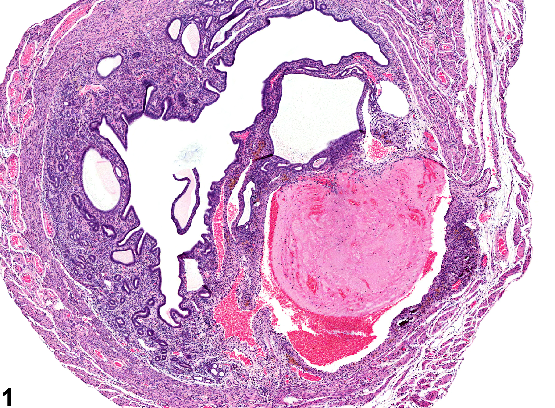
Large or small thrombi may occlude uterine blood vessels in mice (Figure 1 and Figure 2). A distinct endothelial lining may not be observed due to necrosis or obscuring by detritus. This may be associated with prominence and dilation of the surrounding blood vessels (angiectasis). Thrombi must be differentiated from hemangioma and hemangiosarcoma. Angiectasis is characterized by blood vessels lined by a simple nonhypertrophic endothelial lining and is frequently seen with thrombosis. Hemangioma and hemangiosarcoma have plump lining endothelial cells. The endothelial cells of hemangiosarcoma exhibit anaplasia and an increased rate of mitoses.
Uterus - Thrombosis should be diagnosed but need not be graded. Thrombi that are secondary to another process (a neoplasm) should not be diagnosed. Necrosis that is caused by a thrombus should be diagnosed and graded separately.
Davis BJ, Dixon D, Herbert RA. 1999. Ovary, oviduct, uterus, cervix and vagina. In: Pathology of the Mouse: Reference and Atlas (Maronpot RR, Boorman GA, Gaul BW, eds). Cache River Press, Vienna, IL, 409-444.
Maekawa A, Maita K. 1996. Changes in the uterus and vagina. In: Pathobiology of the Aging Mouse (Mohr U, Dungworth DL, Capen CC, Carlton WW, Sundberg JP, Ward JM, eds). ILSI Press, Washington, DC, 469-480.
National Toxicology Program. 1993. NTP TR-428. Toxicology and Carcinogenesis Studies of Manganese (II) Sulfate Monohydrate (CAS No. 10034-96-5) in F344/N Rats and B6C3F1 Mice (Feed Studies). NTP, Research Triangle Park, NC.
Abstract: https://ntp.niehs.nih.gov/go/6000
Uterus - Thrombus in a female B6C3F1/N mouse from a chronic study. A large thrombus is evident in the myometrium.


