Alimentary System
Esophagus - Ulcer
Narrative
Comment:
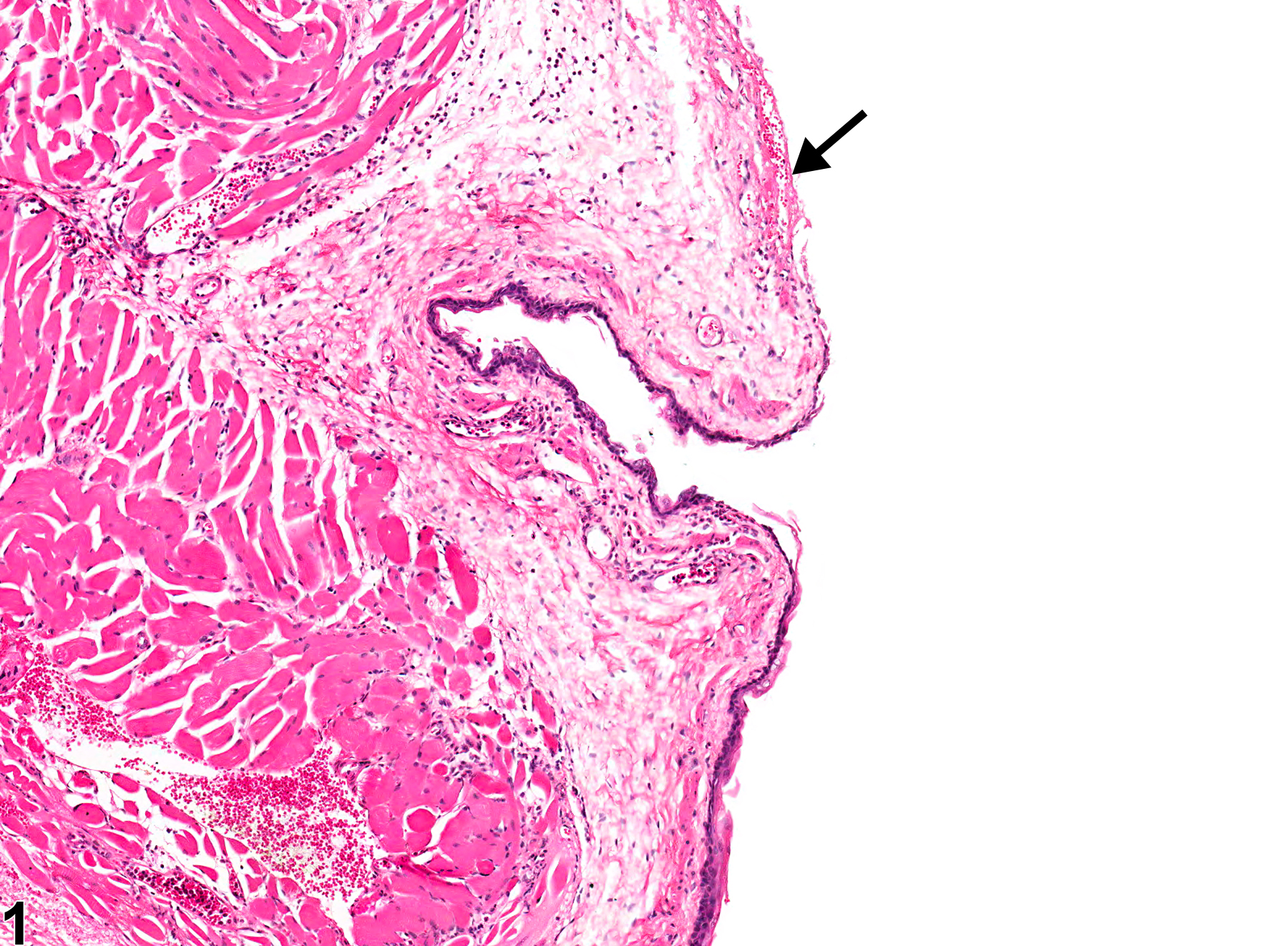
Esophageal erosions, ulcers, and necrosis are frequently the result of gavage accidents. Such trauma can lead to perforation, periesophageal abscesses, and fibrosis. An erosion is defined as loss of the superficial portion (i.e., superficial to the basement membrane) of the tunica mucosa, while an ulcer (Figure 1, Figure 2, Figure 3, Figure 4, and Figure 5, arrows) extends through the basement membrane, exposing the underlying lamina propria. If the overlying epithelium is necrotic but still intact, the lesion is diagnosed as necrosis. Inflammation and regeneration of the epithelium typically accompany ulcerations.
Recommendations:
Whenever present, ulcers should be diagnosed and graded based on the extent of the lesion and number of areas affected. If the epithelial lesion does not extend to the basement membrane, erosion should be diagnosed. If both erosion and ulcer are present concurrently as part of the same lesion, only ulcer is diagnosed. Mucosal epithelial hyperplasia at the edges of an ulcer and inflammation associated with an ulcer are not ordinarily diagnosed unless they are a prominent component of the lesion. It should be noted in the narrative whether the pathologist considers this lesion due to chemical exposure or a gavage error.
References:
Bertram TA, Markovits JE, Juliana MM. 1996. Non-proliferative lesions of the alimentary canal in rats GI-1. In Guides for Toxicologic Pathology. STP/ARP/AFIP, Washington, DC, 1-16.
Full Text: https://www.toxpath.org/docs/SSNDC/GINonproliferativeRat.pdf
Esophagus - Ulcer in a female F344/N rat from a chronic study. A portion of the epithelium is missing (arrow).


