Immune System

Thymus - Atrophy

    Narrative
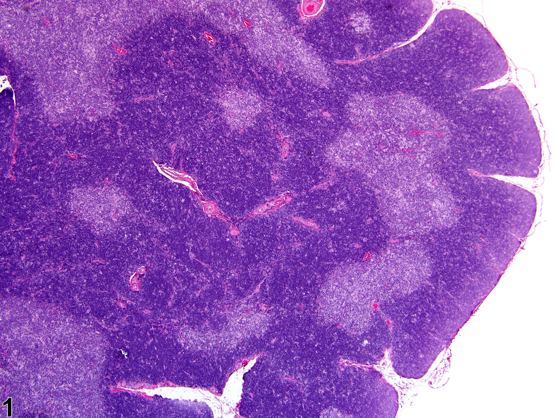
    Thymus - Normal in a male Harlan Sprague-Dawley rat from a subchronic study. The ratio of cortex to medulla is approximately 2:1 (1:1:1, two cortices to medulla).
    Thymus - Normal in a male Harlan Sprague-Dawley rat from a subchronic study. The ratio of cortex to medulla is approximately 2:1 (1:1:1, two cortices to medulla).
    Thymus - Normal in a male Harlan Sprague-Dawley rat from a subchronic study (higher magnification of Figure 1). The lymphocytes are more numerous in the cortex than in the medulla.
    Thymus - Normal in a male Harlan Sprague-Dawley rat from a subchronic study (higher magnification of Figure 1). The lymphocytes are more numerous in the cortex than in the medulla.
    Thymus - Atrophy in a treated female F344/NTac rat from a subchronic study. The cortex, showing minimal atrophy, is thinner and more irregular compared with normal (Figure 1).
    Thymus - Atrophy in a treated female F344/NTac rat from a subchronic study. The cortex, showing minimal atrophy, is thinner and more irregular compared with normal (Figure 1).
    Thymus - Atrophy in a treated female F344/NTac rat from a subchronic study (higher magnification of Figure 3). The cortex, showing minimal atrophy, is thinner and more irregular compared with normal (Figure 2).
    Thymus - Atrophy in a treated female F344/NTac rat from a subchronic study (higher magnification of Figure 3). The cortex, showing minimal atrophy, is thinner and more irregular compared with normal (Figure 2).
    Thymus - Atrophy in a treated female Harlan Sprague-Dawley rat from a chronic study. With mild atrophy, the cortex becomes progressively thinner and the cortical-medullary junction becomes less distinct.
    Thymus - Atrophy in a treated female Harlan Sprague-Dawley rat from a chronic study. With mild atrophy, the cortex becomes progressively thinner and the cortical-medullary junction becomes less distinct.
    Thymus - Atrophy in a treated female Harlan Sprague-Dawley rat from a chronic study (higher magnification of Figure 5). The cortex is thinner, and the cortical-medullary junction is less distinct compared with Figure 4.
    Thymus - Atrophy in a treated female Harlan Sprague-Dawley rat from a chronic study (higher magnification of Figure 5). The cortex is thinner, and the cortical-medullary junction is less distinct compared with Figure 4.
    Thymus - Atrophy in a treated female Harlan Sprague Dawley from a chronic study. With moderate atrophy, delineation of the cortex and medulla is multifocally indistinct.
    Thymus - Atrophy in a treated female Harlan Sprague Dawley from a chronic study. With moderate atrophy, delineation of the cortex and medulla is multifocally indistinct.
    Thymus - Atrophy in a treated female Harlan Sprague Dawley from a chronic study (higher magnification of Figure 7). With moderate atrophy, delineation of the cortex and medulla is multifocally indistinct.
    Thymus - Atrophy in a treated female Harlan Sprague Dawley from a chronic study (higher magnification of Figure 7). With moderate atrophy, delineation of the cortex and medulla is multifocally indistinct.
    Thymus - Atrophy in a treated female Harlan Sprague-Dawley from a chronic study. With marked atrophy, the lack of distinction between the cortex and medulla due to lymphocyte depletion gives the thymus a more uniform appearance.
    Thymus - Atrophy in a treated female Harlan Sprague-Dawley from a chronic study. With marked atrophy, the lack of distinction between the cortex and medulla due to lymphocyte depletion gives the thymus a more uniform appearance.
    Thymus - Atrophy in a treated female Harlan Sprague-Dawley from a chronic study (higher magnification of Figure 9). With marked atrophy, the distinction between the thymic cortex and medulla is no longer visible due to lymphocyte depletion.
    Thymus - Atrophy in a treated female Harlan Sprague-Dawley from a chronic study (higher magnification of Figure 9). With marked atrophy, the distinction between the thymic cortex and medulla is no longer visible due to lymphocyte depletion.

    Authors

    Kristen Hobbie, DVM, PhD
    Principal Pathologist
    Huntingdon Life Sciences
    Peterborough, UK

    Susan A. Elmore, MS, DVM, DACVP, DABT, FIATP
    Staff Scientist, NTP Pathologist
    NTP Pathology Group
    National Toxicology Program
    National Institute of Environmental Health Sciences
    Research Triangle Park, NC

    Holly M. Kolenda-Roberts, DVM, PhD, DACVP
    Veterinary Pathologist
    SNBL USA
    Everett, WA

    Reviewers

    Jerrold M. Ward, DVM, PhD, DACVP, FIATP
    Veterinary Pathologist
    Global VetPathology
    Montgomery Village, MD

    Robert R. Maronpot, DVM, MS, MPH, DACVP, DABT, FIATP
    Senior Pathologist
    Experimental Pathology Laboratories, Inc.
    Research Triangle Park, NC