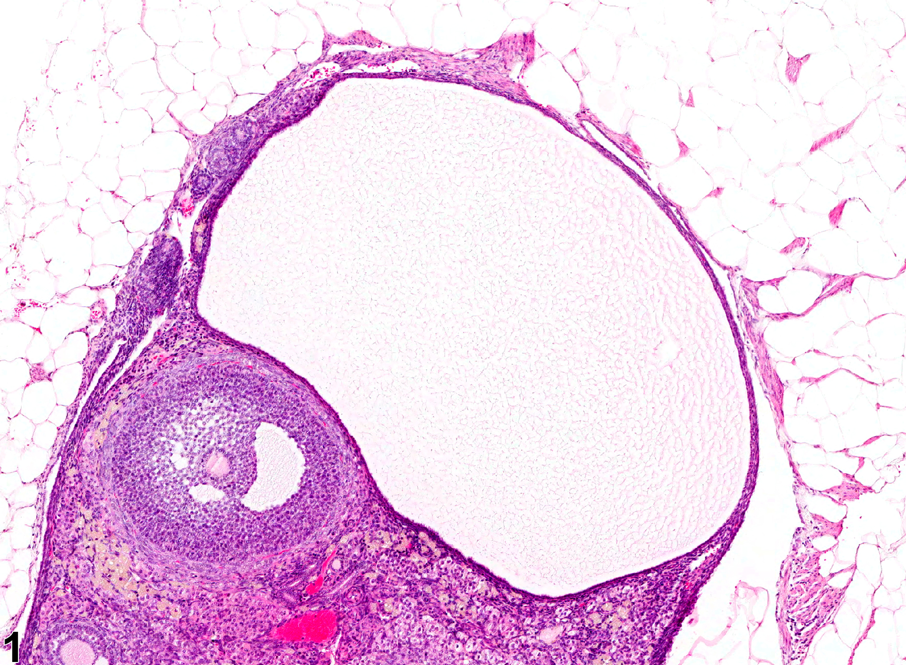
Reproductive System, Female
Ovary, Follicle - Cyst
Narrative
Ovarian follicular cysts are common aging findings in rats and mice. They arise in secondary follicles that fail to ovulate. They vary in size but are larger than a normal preovulatory follicle and may be single or multiple, unilateral or bilateral. Their numbers usually increase with aging. They have also been induced by chemicals that modify cyclical ovarian activity. They may be associated with persistent estrus, cystic endometrial hyperplasia, and infertility. Histologically these cysts are thin walled and filled with pale acidophilic residue or blood; they may contain cell debris, degenerating oocytes, or foamy, vacuolated, or pigment-laden macrophages. These follicles are lined by one to four layers of cuboidal granulosa cells, and there is no luteinization. Some larger cysts may be lined by a single layer of flattened cells resting on a thin fibrous capsule.
Ovarian follicular cysts should be diagnosed, but cysts occurring as background lesions need not be graded. In the case of a follicular cyst, the diagnosis should include the type of cyst/location as a modifier (i.e., Ovary, Follicle - Cyst). If the cysts are thought to be treatment related, they may be graded to fully characterize the treatment effect. If applicable, the terms "bilateral" and "multiple" may be included in the diagnosis.
Alison RH, Morgan KT, Montgomery CA. 1990. Ovary. In: Pathology of the Fischer Rat: Reference and Atlas (Boorman GA, Eustis SL, Elwell MR, Montgomery CA, MacKenzie WF, eds). Academic Press, San Diego, CA, 429-442.
Davis BJ, Dixon D, Herbert RA. 1999. Ovary, oviduct, uterus, cervix and vagina. In: Pathology of the Mouse: Reference and Atlas (Maronpot RR, Boorman GA, Gaul BW, eds). Cache River Press, Vienna, IL, 409-444.
Dixon D, Alison R, Bach U, Colman K, Foley GL, Harleman JH, Hawarth R, Herbert R, Heuser A, Long G, Mirsky M, Regan K, Van Esch E, Westwood FR, Vidal J, Yoshida M. 2014. Nonproliferative and proliferative lesions of the rat and mouse female reproductive system (INHAND). J Toxicol Pathol 27(suppl):1S-107S.
Full Text: https://www.ncbi.nlm.nih.gov/pmc/articles/PMC4253081/Dixon D, Heider K, Elwell MR. 2001. Incidence of nonneoplastic lesions in historical control male and female Fischer-344 rats from 90-day toxicity studies. Toxicol Pathol 23:338-348.
Abstract: https://www.ncbi.nlm.nih.gov/pubmed/7659956Greaves P. 2012. Female genital tract. In: Histopathology of Preclinical Toxicity Studies: Interpretation and Relevance in Drug Safety Evaluation, 4th ed. Elsevier, Amsterdam, 667-724.
Maekawa A, Maita K, Harleman JH. 1996. Changes in the ovary. In: Pathobiology of the Aging Mouse (Mohr U, Dungworth DL, Capen CC, Carlton WW, Sundberg JP, Ward JM, eds). ILSI Press, Washington, DC, 451-467.
Maekawa A, Yoshida A. 1996. Susceptibility of the female genital system to toxic substances. In: Pathobiology of the Aging Mouse, Vol 1 (Mohr U, Dungworth DL, Capen CC, Carlton WW, Sundberg JP, Ward JM, eds). ILSI Press, Washington, DC, 481-493.
Montgomery CA, Alison RH. 1987. Non-neoplastic lesions of the ovary in Fischer 344 rats and B6C3F1 mice. Environ Health Perspect 73:53-75.
Abstract: https://www.ncbi.nlm.nih.gov/pmc/articles/PMC1474552/National Toxicology Program. 2000. NTP TR-476. Toxicology and Carcinogenesis Studies of Primidone (CAS No. 125-33-7) in F344/N Rats and B6C3F1 Mice (Feed Studies). NTP, Research Triangle Park, NC.
Abstract: https://ntp.niehs.nih.gov/go/9754Peluso JJ, Gordon LR. 1992. Nonneoplastic and neoplastic changes in the ovary. In: Pathobiology of the Aging Rat (Mohr U, Dungworth DL, Capen CC, eds). ILSI Press, Washington, DC, 351-364.

Ovary, Follicle - Cyst in a female B6C3F1/N mouse from a chronic study. A large ovarian follicular cyst appears as a distended fluid-filled space lacking an oocyte.


