Respiratory System

Lung, Alveolar/Bronchiolar Epithelium - Hyperplasia

    Narrative
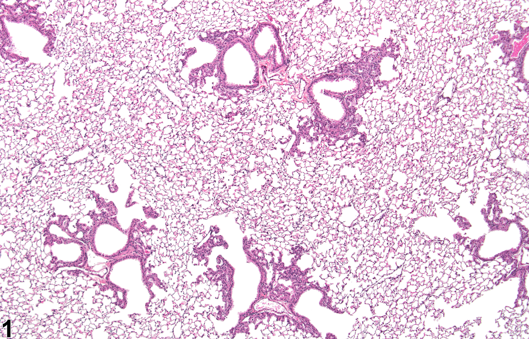
    Lung, Alveolar/bronchiolar epithelium - Hyperplasia in a male B6C3F1/N mouse from a chronic study. This low-magnification image shows the distribution of the lesion.
    Lung, Alveolar/bronchiolar epithelium - Hyperplasia in a male B6C3F1/N mouse from a chronic study. This low-magnification image shows the distribution of the lesion.
    Lung, Alveolar/bronchiolar epithelium - Hyperplasia in a male B6C3F1/N mouse from a chronic study. This lesion occurs adjacent to bronchioles and alveolar ducts.
    Lung, Alveolar/bronchiolar epithelium - Hyperplasia in a male B6C3F1/N mouse from a chronic study. This lesion occurs adjacent to bronchioles and alveolar ducts.
    Lung, Alveolar/bronchiolar epithelium - Hyperplasia in a male B6C3F1/N mouse from a chronic study (higher magnification of Figure 2). The majority of the cells are not ciliated, but some are (arrows).
    Lung, Alveolar/bronchiolar epithelium - Hyperplasia in a male B6C3F1/N mouse from a chronic study (higher magnification of Figure 2). The majority of the cells are not ciliated, but some are (arrows).
    Lung, Alveolar/bronchiolar epithelium - Hyperplasia in a male B6C3F1/N mouse from a chronic study. Secondary alveolar epithelial hyperplasia associated with inflammation is present at the bronchiolar-alveolar duct junction.
    Lung, Alveolar/bronchiolar epithelium - Hyperplasia in a male B6C3F1/N mouse from a chronic study. Secondary alveolar epithelial hyperplasia associated with inflammation is present at the bronchiolar-alveolar duct junction.
    Lung, Alveolar epithelium - Hyperplasia in a male B6C3F1/N mouse from a chronic study (higher magnification of Figure 4). Large, foamy macrophages, neutrophils, and lymphocytes are associated with the alveolar epithelial hyperplasia.
    Lung, Alveolar epithelium - Hyperplasia in a male B6C3F1/N mouse from a chronic study (higher magnification of Figure 4). Large, foamy macrophages, neutrophils, and lymphocytes are associated with the alveolar epithelial hyperplasia.

    Authors

    Mark F. Cesta, DVM, PhD, DACVP
    Staff Scientist/NTP Pathologist
    NTP Pathology Group
    National Toxicology Program
    National Institute of Environmental Health Sciences
    Research Triangle Park, NC


    Darlene Dixon, DVM, PhD, DACVP
    Group Leader
    Molecular Pathogenesis Group
    National Toxicology Program
    National Institute of Environmental Health Sciences
    Research Triangle Park, NC


    Ronald A. Herbert, DVM, PhD
    Group Leader/NTP Pathologist
    Pathology Support Group
    National Toxicology Program
    National Institute of Environmental Health Sciences
    Research Triangle Park, NC


    Lauren M. Staska, DVM, PhD, DACVP
    Senior Pathologist
    WIL Research
    Hillsborough, NC

    Reviewers

    Gary A. Boorman, DVM, PhD, DACVP, DABT, DACLAM
    Pathologist
    Covance Laboratories, Inc.
    Chantilly, VA

    Robert C. Sills, DVM, PhD, FIATP
    Chief, Cellular and Molecular Pathology Branch
    National Toxicology Program
    National Institute of Environmental Health Sciences, Research Triangle Park, NC