Reproductive System, Female
Uterus - Decidual Reaction
Narrative
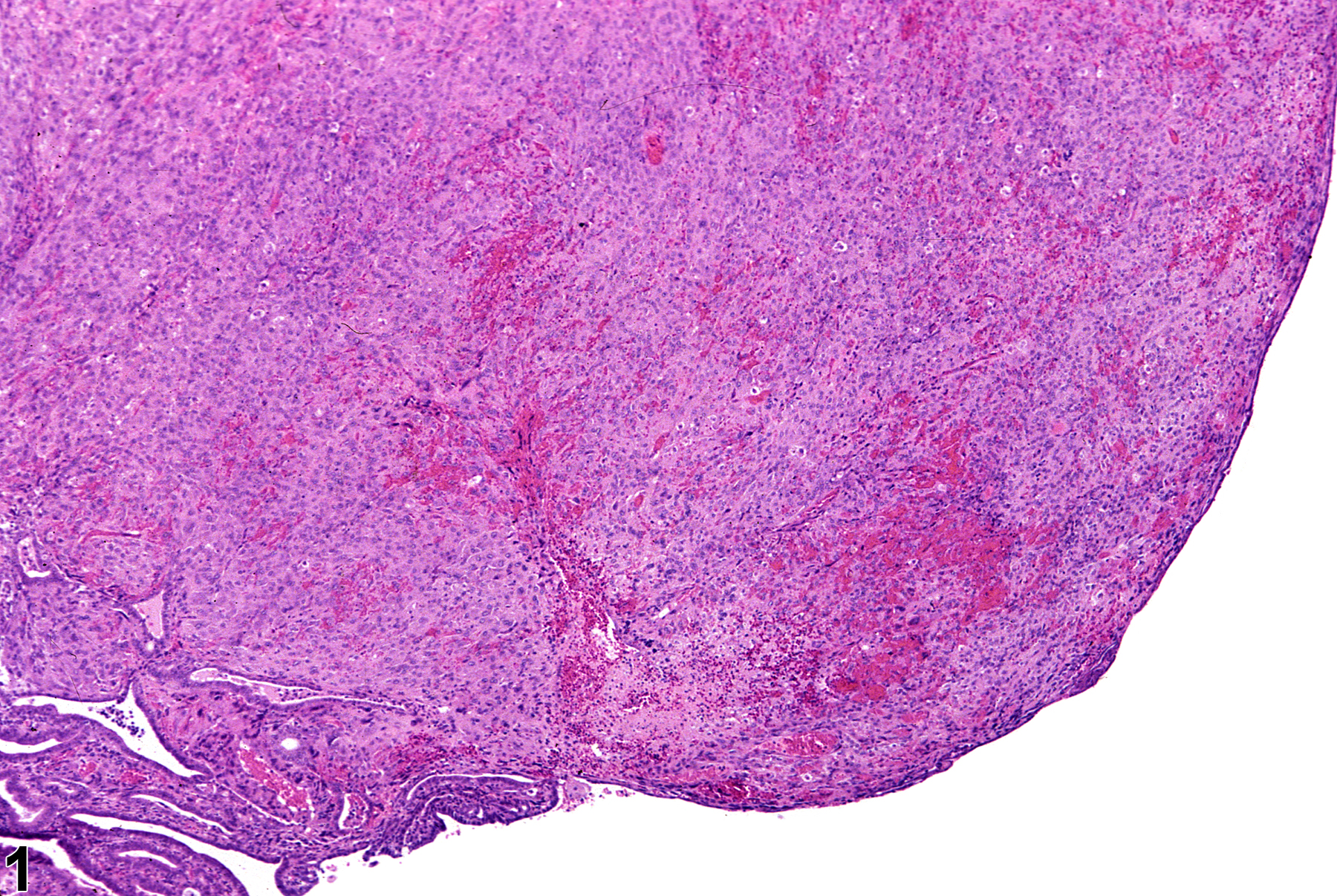
Decidual reactions (Figure 1, Figure 2, Figure 3, and Figure 4) are focal, nonneoplastic, proliferative changes in the endometrium. They are observed as localized nodules consisting of hypertrophied stromal cells with epithelioid transformation and, sometimes, bizarre nuclei resembling the trophoblasts observed in mature placentae. There is no encapsulation, and clear boundaries around the nodules are not apparent. Decidual reactions are characterized by large rounded stromal cells with abundant eosinophilic cytoplasm, large round nuclei, and prominent nucleoli and occasional multinucleated cells. This lesion has been referred to as a deciduoma of decidual alteration.
The pathologist needs to be aware of the uniqueness of decidual reactions so they will not be confused with neoplasia. This proliferative response mimics normal implantation sites and may occur in young and old, nonpregnant or pseudopregnant rats and is occasionally found in 90-day studies and in aged virgin mice. Decidual reactions appear as discrete round nodules in the uterine horn. They may be single or multiple, unilateral or bilateral, and a focal lesion of decidual alteration may occur in a polyp or neoplasms. They can be induced in rats by growth hormone and by a number of agents, such as intrauterine instillation of sesame oil, prostaglandin E2, or progestins. Decidual reactions can also be induced by mechanical stimulation or irritation, endometrial traumatization, and intrauterine injection of a variety of substances (balanced salt, oily fluids, and air).
Brown RH, Leininger JR. 1992. Alterations of the uterus. In: Pathobiology of the Aging Rat (Mohr U, Dungworth DL, Capen CC, eds). ILSI Press, Washington, DC, 377–388.
Davis BJ, Dixon D, Herbert RA. 1999. Ovary, oviduct, uterus, cervix and vagina. In: Pathology of the Mouse: Reference and Atlas (Maronpot RR, Boorman GA, Gaul BW, eds). Cache River Press, Vienna, IL, 409-444.
Dixon D, Heider K, Elwell M. 1995. Incidence of nonneoplastic lesions in historical control male and female Fischer 344 rats from 90 day toxicity studies. Toxicol Pathol 23:338–348.
Abstract: https://www.ncbi.nlm.nih.gov/pubmed/7659956Gopinath C, Prentice DE, Lewis DJ. 1987. The reproductive system. In: Atlas of Experimental Toxicology (Gresham GA, eds). MTP Press, Lancaster, UK, 91-103.
Greaves P. 2012. Female genital tract. In: Histopathology of Preclinical Toxicity Studies: Interpretation and Relevance in Drug Safety Evaluation, 4th ed. Elsevier, Amsterdam, 667-724.
Leininger JR, Jokinen MP. 1990. Oviduct, uterus and vagina. In: Pathology of the Fischer Rat (Boorman GA, Eustis SL, Elwell MR, Montgomery CA, MacKenzie WF, eds). Academic Press, San Diego, CA, 443-459.
Maekawa A, Maita K. 1996. Changes in the uterus and vagina. In: Pathobiology of the Aging Mouse (Mohr U, Dungworth DL, Capen CC, Carlton WW, Sundberg JP, Ward JM, eds). ILSI Press, Washington, DC, 469-480.
National Toxicology Program. 1988. NTP TR-334. Toxicology and Carcinogenesis Studies of 2-Amino-5-nitrophenol (CAS No. 121-88-0) in F344/N Rats and B6C3F1 Mice (Gavage Studies). NTP, Research Triangle Park, NC.
Abstract: https://ntp.niehs.nih.gov/go/8904
Uterus - Decidual reaction in a female F344/N rat from a chronic study. There is an area of decidual reaction in the uterus.


