Endocrine System
Pituitary Gland, Pars Distalis - Necrosis
Narrative
Carroll R. 1967. The pathogenesis of pituitary and adrenal necrosis. Ir J Med Sci 6:453-464.
Abstract: http://link.springer.com/article/10.1007/BF02954153Daniel PM, Prichard MM. 1956. Anterior pituitary necrosis; infarction of the pars distalis produced experimentally in the rat. Q J Exp Physiol Cogn Med Sci 41:215-229.
Full Text: http://ep.physoc.org/content/41/3/215.longSert M, Tetiker T, Kirim S, Kocak M. 2003. Clinical report of 28 patients with Sheehan's syndrome. Endocr J 50:297-301.
Abstract: http://www.ncbi.nlm.nih.gov/pubmed/12940458
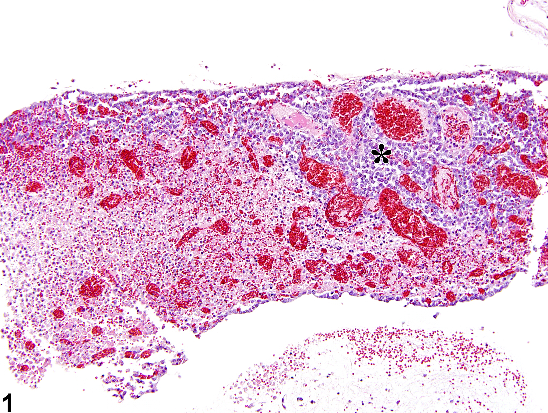
Pituitary Gland, Pars distalis - Necrosis in a female R344/N rat from a subchronic study. Loss of cellular detail is present in the area of necrosis with normal pituitary present in the upper right (asterisk); vascular congestion is present in both areas.


