Reproductive System, Female
Vagina - Angiectasis
Narrative
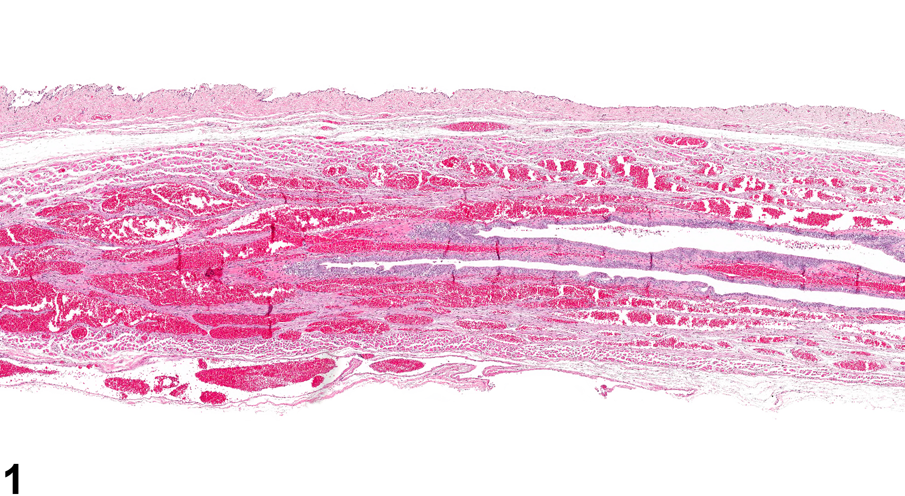
Dilated vessels in the rat vagina are rarely observed. When present as angiectasis, they are usually a collection of prominent dilated blood vessels lined by a single layer of endothelium with flattened nuclei (Figure 1 and Figure 2). A distinction between angiectasis and hemangioma should be attempted, although the distinction is not always obvious. Hemangiomas tend to be well-circumscribed, unencapsulated masses composed of tightly packed, dilated vascular spaces. Each vascular space is enclosed and lined by a single layer of normal-appearing endothelial cells aligned on collagenous septa, which are usually thin, although some have broad collagenous stroma. In contrast, angiectasis does not usually present a well-circumscribed mass: the dilated vascular channels course irregularly through the tissue.
National Toxicology Program. 1986. NTP TR-250. Toxicology and Carcinogenesis Studies of Benzyl Acetate (CAS No. 140-11-4) in F344/N Rats and B6C3F1 Mice (Gavage Studies). NTP, Research Triangle Park, NC.
Abstract: https://ntp.niehs.nih.gov/go/7108National Toxicology Program. 1993. NTP TR-431. Toxicology and Carcinogenesis Studies of Benzyl Acetate (CAS No. 140-11-4) in F344/N Rats and B6C3F1 Mice (Feed Studies). NTP, Research Triangle Park, NC.
Abstract: https://ntp.niehs.nih.gov/go/6006
Vagina - Angiectasis in a female F344/N rat from a chronic study. Dilated blood vessels are present in the wall of the vagina.


