Musculoskeletal System
Bone - Fracture
Narrative
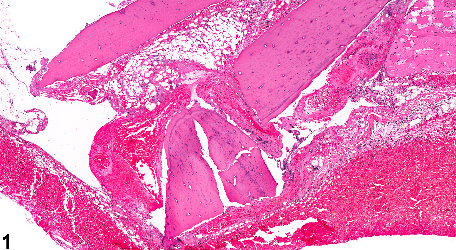
Bone fractures (Figure 1 and Figure 2) are characterized by discontinuity of cortical bone and/or disruption of medullary trabeculae (microfracture). They are often associated with variable hemorrhage, inflammation, and fibrosis, depending on the severity and chronicity of the lesion. In acute fractures large amounts of hemorrhage are common, and with chronicity increased numbers of inflammatory cells and fibrous tissue response are elicited, with eventual organized fibrosis, bony remodeling, and repair through callus formation.
Fractures may be secondary to severe inflammatory, neoplastic, or metabolic disease. Fractures have been also noted secondary to bone disease due to treatment with some compounds. For example, excessive vitamin A treatment leads to decreased bone formation and degenerative physeal lesions in rats, which result in decreased longitudinal bone growth.
Occasionally, sectioning artifacts can appear as fractures and must be differentiated from true fractures. Artifactual fractures, or pseudofractures, will lack the hemorrhage, fibrin deposition, fibrosis, and callus formation that might be seen with a true fracture.
Leininger JR, Riley MGI. 1990. Bones, joints, and synovia. In: Pathology of the Fischer Rat: Reference and Atlas (Boorman G, Eustis SL, Elwell MR, Montgomery CA, MacKenzie WF, eds). Academic Press, San Diego, 209-226.
Long PH, Leininger JR. 1999. Bones, joints, and synovia. In: Pathology of the Mouse (Maronpot R, Boorman G, Gaul BW, eds). Cache River Press, St Louis, 645-678.

Bone - Fracture in a female F344/N rat. A section of tibia illustrates a fracture of cortical bone with displacement of the distal tibia and associated hemorrhage and fibrin deposition.


