Reproductive System, Female
Uterus - Mesonephric Duct Remnant
Narrative
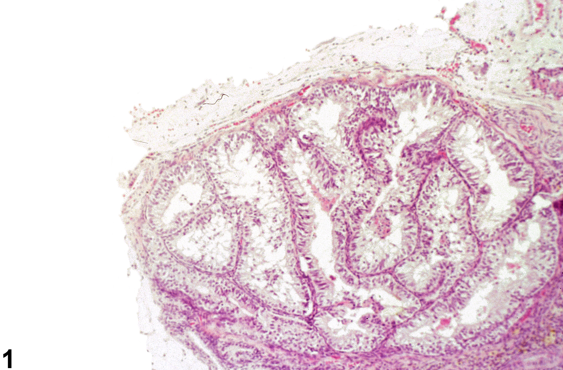
Remnants of the mesonephric duct (Figure 1) appear as coiled or branching tubules lined by pseudostratified columnar epithelium and smooth muscle and are reminiscent of that seen in the ductus epididymides and vas deferens. Typically these structures are seen laterally attached to the uterine horns or body, and they may be unilateral or bilateral. They are typically present at the site of attachment of the broad ligament of the uterus. These embryonic remnants must be differentiated from neoplasms, which display growth patterns characteristic of the tumor type.
Uterus - Mesonephric duct remnant should be diagnosed but need not be graded unless grading would help characterize a treatment effect.
Davis BJ, Dixon D, Herbert RA. 1999. Ovary, oviduct, uterus, cervix and vagina. In: Pathology of the Mouse: Reference and Atlas (Maronpot RR, Boorman GA, Gaul BW, eds). Cache River Press, Vienna, IL, 409-444.
Dixon D, Alison R, Bach U, Colman K, Foley GL, Harleman JH, Hawarth R, Herbert R, Heuser A, Long G, Mirsky M, Regan K, Van Esch E, Westwood FR, Vidal J, Yoshida M. 2014. Nonproliferative and proliferative lesions of the rat and mouse female reproductive system (INHAND). J Toxicol Pathol 27(suppl):1S-107S.
Full Text: https://www.ncbi.nlm.nih.gov/pmc/articles/PMC4253081/Leininger JR, Jokinen MP. 1990. Oviduct, uterus and vagina. In: Pathology of the Fischer Rat (Boorman GA, Eustis SL, Elwell MR, Montgomery CA, MacKenzie WF, eds). Academic Press, San Diego, CA, 443-459.
National Toxicology Program. 1990. NTP TR-376. Toxicology and Carcinogenesis Studies of Allyl Glycidyl Ether (CAS No. 106-92-3) in Osborne-Mendel Rats and B6C3F1 Mice (Inhalation Studies). NTP, Research Triangle Park, NC.
Abstract: https://ntp.niehs.nih.gov/go/8892
Uterus - Mesonephric duct remnant in a female Osborne Mendel rat from a chronic study. Tubules lined by pseudostratified columnar epithelium and smooth muscle are adjacent to the uterus.


