Reproductive System, Female
Ovary - Pigment
Narrative
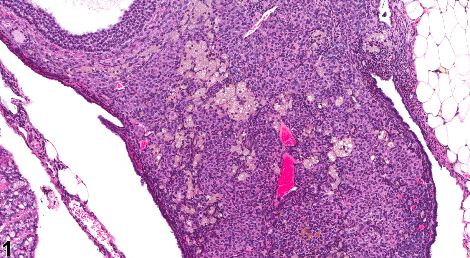
Various pigments (Figure 1, Figure 2, Figure 3 and Figure 4) seen in ovaries include ceroid, hemosiderin, hematoidin, and melanin. In the NTP program, yellow-brown, periodic-acid-Schiff-positive and acid-fast ceroid pigment has been observed in Fischer 344 rats and B6C3F1 mice but is more common in mice. Yellow to brown pigment is often seen as an aging change in mouse and rat ovaries. Ceroid is the pigment most frequently seen, and this accumulates mainly in the cytoplasm of interstitial cells. Administration of compounds that interfere with steroid synthesis may result in accumulation of lipofuscin. Accumulation of hemosiderin and hematoidin may be associated with vascular lesions resulting in hemorrhage into follicles or corpora lutea. Accumulation of melanin has been reported in the ovaries of pigmented B6C3F1 mice, but the mechanism involved has not been clarified.
Ovary - Pigment should be diagnosed when it is considered to be relevant to the study (or if it is unclear whether it is relevant to the study) or if it is unusually prominent. Not all pigments have to be diagnosed, as some are ubiquitous in aging animals or are secondary to another disease process (e.g., hemorrhage) and not toxicologically meaningful. The pathologist should use his or her judgment in deciding whether pigment should be diagnosed. Definitive pigment identification is often difficult in histologic sections, even with a battery of special stains. Therefore, a diagnosis of "pigment" (as opposed to diagnosing the type of pigment, e.g., hemosiderin or lipofuscin) is most appropriate. The pathology narrative should describe the morphologic features of the pigmentation. When pigment is diagnosed, it should be given a severity grade.
Alison RH, Morgan KT, Montgomery CA. 1990. Ovary. In: Pathology of the Fischer Rat: Reference and Atlas (Boorman GA, Eustis SL, Elwell MR, Montgomery CA, MacKenzie WF, eds). Academic Press, San Diego, CA, 429-442.
Davis BJ, Dixon D, Herbert RA. 1999. Ovary, oviduct, uterus, cervix and vagina. In: Pathology of the Mouse: Reference and Atlas (Maronpot RR, Boorman GA, Gaul BW, eds). Cache River Press, Vienna, IL, 409-444.
Maekawa A, Maita K, Harleman JH. 1996. Changes in the ovary. In: Pathobiology of the Aging Mouse (Mohr U, Dungworth DL, Capen CC, Carlton WW, Sundberg JP, Ward JM, eds). ILSI Press, Washington, DC, 451-467.
Montgomery CA, Alison RH. 1987. Non-neoplastic lesions of the ovary in Fischer 344 rats and B6C3F1 mice. Environ Health Perspect 73:53-75.
Abstract: https://www.ncbi.nlm.nih.gov/pmc/articles/PMC1474552/National Toxicology Program. 2001. NTP TR-497. Toxicology and Carcinogenesis Studies of Methacrylonitrile (CAS No. 126-98-7) in F344/N Rats and B6C3F1 Mice (Gavage Studies). NTP, Research Triangle Park, NC.
Abstract: https://ntp.niehs.nih.gov/go/14872
Ovary - Pigment in a female B6C3F1/N mouse from a chronic study. Scattered foci of pigment are present throughout ovarian parenchyma.


