Reproductive System, Female
Ovary - Necrosis
Narrative
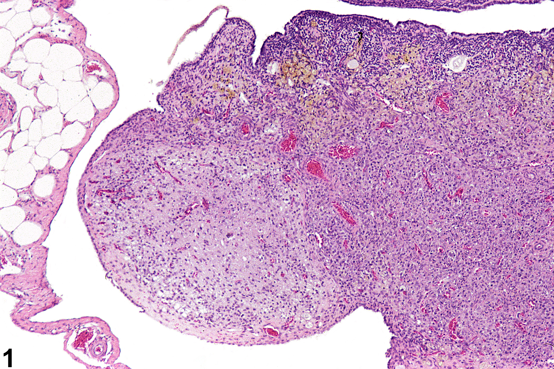
Ovary - Necrosis may be present is a well-circumscribed, unencapsulated area composed of amorphous necrotic cellular debris centrally and moderate numbers of mainly mononuclear inflammatory cells peripherally (Figure 1, Figure 2 and Figure 3). The features of necrosis in the ovary, as in other tissues, include nuclear pyknosis and/or karyorrhexis, cytoplasmic eosinophilia, and cellular swelling or shrinkage.
Ovary - Necrosis should be diagnosed and graded when it is a primary lesion. If the necrosis is secondary to another lesion, such as inflammation, it should not be diagnosed separately unless warranted by severity but should be described in the pathology narrative. Lesions that are considered secondary to the necrosis, such as inflammation or hemorrhage, should not be diagnosed separately unless warranted by severity. Apoptotic cells in atretic follicles or regressing corpora lutea should not be diagnosed as necrosis.
Greaves P. 2012. Female genital tract. In: Histopathology of Preclinical Toxicity Studies: Interpretation and Relevance in Drug Safety Evaluation, 4th ed. Elsevier, Amsterdam, 667-724.
National Toxicology Program. 1988. NTP TR-330. Toxicology and Carcinogenesis Studies of 4-Hexylresorcinol (CAS No. 136-77-6) in F344/N Rats and B6C3F1 Mice (Gavage Studies). NTP, Research Triangle Park, NC.
Abstract: https://ntp.niehs.nih.gov/go/8896
Ovary - Necrosis in a female F344/N rat from a chronic study. The necrotic ovarian tissue is sharply demarcated from the adjacent ovary.


