Endocrine System
Adrenal Gland - Accessory Adrenocortical Nodule
Narrative
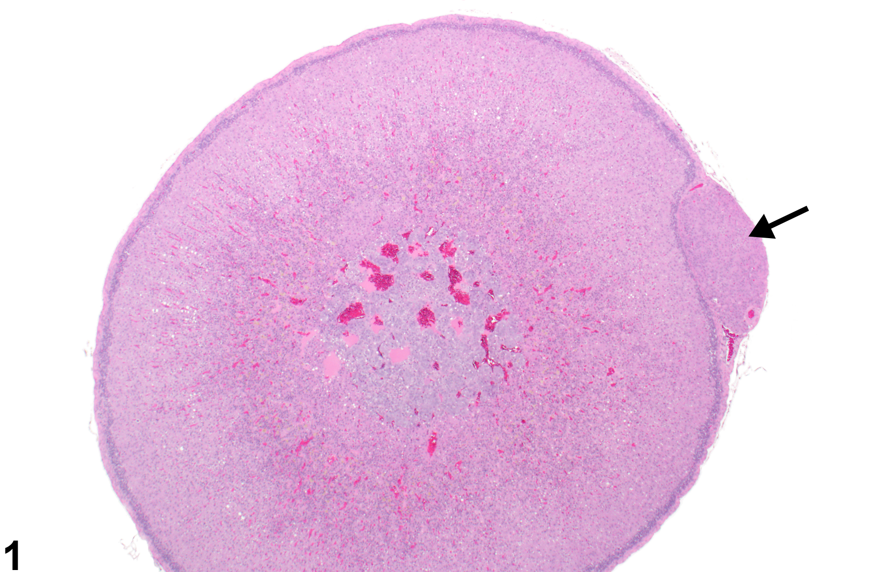
Most nodules are spherical to ovoid structures surrounded by a usually delicate fibrous capsule and composed of well-differentiated zonae glomerulosa-type and fasciculata-type cells (Figure 1 and Figure 2), sometimes admixed and sometimes arranged in typical layers. Some adrenocortical accessory tissue consists of less densely packed nests of more primitive-appearing cortical cells (Figure 3 and Figure 4). The typical surrounding fibrous capsule distinguishes accessory adrenocortical nodules from focal cortical hyperplasia and from extracapsular protrusions of cortical tissue. Accessory nodules must also be distinguished from cortical adenomas and carcinomas, which exhibit cellular pleomorphism and atypia and loss of cortical layering but are not commonly encapsulated.
Dunn TB. 1970. Normal and pathologic anatomy of the adrenal gland of the mouse, including neoplasms. J Natl Cancer Inst 44:1323-1389.
Abstract: http://jnci.oxfordjournals.org/content/44/6/1323.abstractHamlin MH, Banas DA. 1990. Adrenal gland. In: Pathology of the Fischer Rat: Reference and Atlas (Boorman GA, Eustis SL, Elwell MR, Montgomery CA, MacKenzie WF, eds). Academic Press, San Diego, 501-518.
Abstract: https://www.ncbi.nlm.nih.gov/nlmcatalog/9002563McInnes EF. 2011. Wistar and Sprague Dawley rats. In: Background Lesions in Laboratory Animals: A Color Atlas (McInnes EF, ed). Saunders Elsevier, Amsterdam, 16-36.
Abstract: http://www.sciencedirect.com/science/book/9780702035197Parker GA, Valerio MG. 1983. Accessory adrenocortical tissue, rat. In: Monographs on the Pathology of Laboratory Animals: Endocrine System (Jones TC, Mohr U, Hunt RD, eds). Springer, Berlin, 16-17.
Abstract: http://www.springer.com/medicine/pathology/book/978-3-642-64649-2National Toxicology Program. 2007. NTP TR-535. Toxicology and Carcinogenesis Studies of 4- Methylimidazole (CAS No. 822-36-6) in F344/N Rats and B6C3F1 Mice (Feed Studies). NTP, Research Triangle Park, NC.
Abstract: https://ntp.niehs.nih.gov/go/13651National Toxicology Program. 2010. NTP TR-560. Toxicology and Carcinogenesis Studies of Androstenedione (CAS No. 63-05-8) in F344/N Rats and B6C3F1 Mice (Gavage Studies). NTP, Research Triangle Park, NC.
Abstract: https://ntp.niehs.nih.gov/go/33555National Toxicology Program. 2013. NTP TR-578. Toxicology and Carcinogenesis Studies of Gingko biloba Extract in F344/N Rats and B6C3F1 Mice (Gavage Studies). NTP, Research Triangle Park, NC.
Abstract: https://ntp.niehs.nih.gov/go/37193Nyska A, Maronpot RR. 1990. Adrenal gland. In: Pathology of the Mouse: Reference and Atlas (Maronpot RR, Boorman GA, Gaul BW, eds). Cache River Press, Vienna, IL, 509-536.
Abstract: http://www.cacheriverpress.com/books/pathmouse.htmSass B. 1983. Accessory adrenocortical tissue, mouse. In: Monographs on the Pathology of Laboratory Animals: Endocrine System (Jones TC, Mohr U, Hunt RD, eds). Springer, Berlin, 12-15.
Abstract: http://www.springer.com/medicine/pathology/book/978-3-642-64649-2Taylor I. 2011. Mouse. In: Background Lesions in Laboratory Animals: A Color Atlas (McInnes EF, ed). Saunders Elsevier, Amsterdam, 45-72.
Abstract: http://www.sciencedirect.com/science/book/9780702035197
Adrenal gland - Accessory adrenocortical nodule in a male F344/N rat from a chronic study. There is a nodule of adrenal cortical cells on the extracapsular surface (arrow).
All Images

Adrenal gland - Accessory adrenocortical nodule in a male F344/N rat from a chronic study (higher magnification of Figure 1). The cells of this accessory adrenocortical nodule (asterisk) at the extracapsular surface are well-differentiated zona fasciculata cells and are surrounded by a delicate fibrous capsule (arrow).

Periadrenal fat - Accessory adrenocortical nodule in a male B6C3F1/N mouse from a chronic study (higher magnification of Figure 5). Accessory cortical nodule (arrow) is composed of well-differentiated adrenal cortical cells surrounded by a thin fibrous capsule in the periadrenal (retroperitoneal) fat.


