Special Senses System
Eye, Retina - Degeneration
Narrative
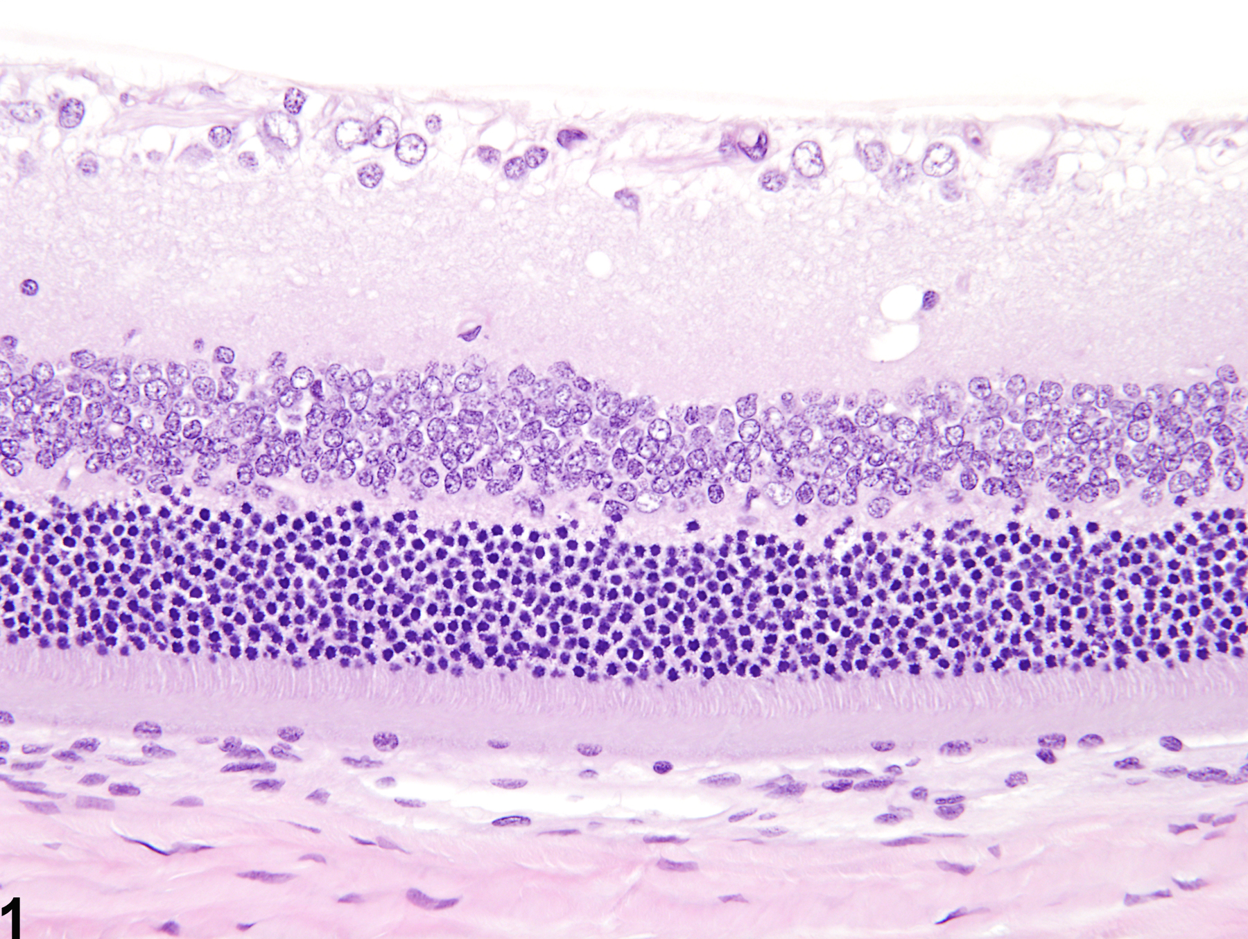
Mild retinal degeneration (Figure 2, compare to Figure 1) typically consists of loss of the rod and cone photoreceptor processes, apoptosis of outer nuclear layer photoreceptor cells, hypocellularity and disorganization of the inner and outer nuclear layers, and narrowing or absence of the plexiform layers. More pronounced retinal degeneration (Figure 3, compare to Figure 1) is typically characterized by overall thinning, architectural disruption, and loss of cellular elements, as well as complete absence of the rod and cone photoreceptors and the plexiform layers. Degenerate retinas may also exhibit other morphologic features, such as diffuse or nodular proliferation of retinal glial cells (e.g., Müller cells and astrocytes), fixed fold and rosette-like formations, and intraretinal cavitations or cysts ("microcystoid" formation and retinoschisis), as well as concurrent proliferation and/or intraretinal migration of the adjacent retinal pigment epithelium.
Rats and mice can also exhibit an incidental aging change known as peripheral retinal degeneration ( and , compare to ), which is characterized by marked thinning of the peripheral retina due to hypocellularity or absence of retinal layers. Although not illustrated, cystic cavitations ("microcystoid" change) are also common features of this peripheral age-related degeneration.
Breider MA, Pilcher GD, Graziano MJ, Gough AW. 1998. Retinal degeneration in rats induced by CI-1010, a 2-nitroimidazole radiosensitizer. Toxicol Pathol 26:234-239.
Full Text: http://tpx.sagepub.com/content/26/2/234.full.pdf+htmlDel Cerro M, Grover D, Monjan A, Pfau C, Dematte J. 1984. Congenital retinitis in the rat following maternal exposure to lymphocytic choriomeningitis virus. Exp Eye Res 38:313-324.
Abstract: https://www.ncbi.nlm.nih.gov/pubmed/6723808De Vera Mudry MC, Kronenberg S, Komatsu S-I, Aguirre GD. 2013. Blinded by the light: Retinal phototoxicity in the context of safety studies. Toxicol Pathol 41: 813-825.
Abstract: https://www.ncbi.nlm.nih.gov/pubmed/23271306DiLoreto DA Jr, Del Cerro C, Cox C, Del Cerro M. 1998. Changes in visually guided behavior of Royal College of Surgeons rats as a function of age: A histologic, morphometric, and functional study. Invest Ophthalmol Vis Sci 39:1058-1063.
Abstract: https://www.ncbi.nlm.nih.gov/pubmed/9579488Frame SR, Carlton WW. 1991. Toxic retinopathy, rat, mouse, and hamster. In: International Life Sciences Institute Monographs on Pathology of Laboratory Animals, Vol 10, Eye and Ear (Jones TC, Mohr U, Hunt RD, eds). Springer, Berlin, 116-124.
Frame SR, Slone TW. 1966. Nonneoplastic and neoplastic changes in the eye. In: Pathobiology of the Aging Mouse, Vol 2 (Mohr U, Dungworth DL, Capen CC, Carlton WW, Sundberg JP, Ward JM, eds). ILSI Press, Washington, DC, 97-103.
Geiss V, Yoshitomi K. 1991. Eyes. In: Pathology of the Mouse: Reference and Atlas (Maronpot RR, Boorman GA, Gaul BW, eds). Cache River Press, Vienna, IL, 471-489.
Abstract: http://www.cacheriverpress.com/books/pathmouse.htmGreaves P. 2007. Nervous system and special sense organs. In: Histopathology of Preclinical Toxicity Studies: Interpretation and Relevance in Drug Safety Evaluation, 3rd ed. Academic Press, San Diego, CA, 861-933.
Abstract: http://www.sciencedirect.com/science/book/9780444527714Illanes O, Anderson S, Niesman M, Zwick L, Jessen BA. 2006. Retinal and peripheral nerve toxicity induced by the administration of a pan-cyclin dependent kinase (cdk) inhibitor in mice. Toxicol Pathol 34:243-248.
Full Text: http://tpx.sagepub.com/content/34/3/243.fullLai Y-L, Jacoby RL, Jonas AM. 1978. Age-related and light-associated retinal changes in Fischer rats. Invest Ophthalmol Vis Sci 17:634-638.
Abstract: https://pubmed.ncbi.nlm.nih.gov/669893/Lee EW, Render JA, Garner CD, Brady AN, Li LC. 1990. Unilateral degeneration of retina and optic nerve in Fischer-344 rats. Vet Pathol 27:439-444.
Abstract: http://vet.sagepub.com/content/27/6/439.shortMecklenburg L, Schraermeyer U. 2007. An overview on the toxic morphological changes in the retinal pigment epithelium after systemic compound administration. Toxicol Pathol 35:252-267.
Full Text: http://tpx.sagepub.com/content/35/2/252.fullMontalbán-Soler L, Alarcón-Martínez L, Jiménez-López M, Salias-Navarro M, Galindo-Romero C, Bezerrade Sá F, García-Ayoso D, Avilés-Trigueros M, Vidal-Sanz M, Agudo-Barrioso M, Villegas-Pérez M. 2012. Retinal compensatory changes after the light damage in albino mice. Mol Vis 18:675-693.
Full Text: https://www.ncbi.nlm.nih.gov/pmc/articles/PMC3325904/National Toxicology Program. 2012. NTP TR-572. Toxicology and Carcinogenesis Studies of Methyl trans-Styryl Ketone (CAS No. 1896-62-4) in F344/N Rats and B6C3F1 Mice (Feed and Dermal Studies). NTP, Research Triangle Park, NC.
Abstract: https://ntp.niehs.nih.gov/go/36154Nyska A, Maronpot RR, Ghanayem BI. 1999. Ocular thrombosis and retinal degeneration induced in female F344 rats by 2-butoxyethanol. Hum Exp Toxicol 18:577-582.
Abstract: http://het.sagepub.com/content/18/9/577.abstractRamos M, Reilly CM, Bolon B. 2011. Toxicological pathology of the retina and optic nerve. In: Fundamental Neuropathology for Pathologists and Toxicologists (Bolon B, Butt MT, eds). Wiley, Hoboken, NJ, 385-412.
Abstract: http://onlinelibrary.wiley.com/doi/10.1002/9780470939956.ch24/summaryRao GN. 1991. Light intensity-associated eye lesions of Fischer 344 rats in long-term studies. Toxicol Pathol 19:148-155.
Abstract: http://tpx.sagepub.com/content/19/2/148.shortSeoane A, Espejo M, Pallàs M, Rodriguez-Farre E, Ambrosio S, Llorens J. 1999. Degeneration and gliosis in rat retina and central nervous system following 3,3'-iminodipropionitrile exposure. Brain Res 833:258-271.
Full Text: http://toxsci.oxfordjournals.org/content/88/2/456.full.pdfSerfilippi LM, Stackhouse Pallman DR, Gruebbel MM, Kern TJ, Spainhour CB. 2004. Assessment of retinal degeneration in outbred albino mice. Comp Med 54:69-76.
Abstract: https://www.ncbi.nlm.nih.gov/pubmed/15027621Smith RS, Hawes NL, Chang B, Nishina PM. 2002. Retina. In: Systematic Evaluation of the Mouse Eye: Anatomy, Pathology, and Biomethods (Smith RS, John SWM, Nishina PM, Sundberg JP, eds). CRC Press, Boca Raton, FL, 195-225.
Sun H, Wang Y, Pang I-H, Shen J, Tang X, Li Y, Liu C, Li B. 2011. Protective effect of a JNK inhibitor against retinal ganglion cell loss induced by acute moderate ocular hypertension. Mol Vis 17:864-875.
Full Text: https://www.ncbi.nlm.nih.gov/pmc/articles/PMC3081797/Tanito M, Kaidzu S, Ohira A, Anderson RE. 2008. Topography of retinal damage in light-exposed albino rats. Exp Eye Res 87:292-295.
Abstract: https://www.ncbi.nlm.nih.gov/pubmed/18586030Taradach C, Greaves P, Rubin LF. 1984. Spontaneous eye lesions in laboratory animals: Incidence in relation to age. Crit Rev Toxicol 12:121-147.
Abstract: https://www.ncbi.nlm.nih.gov/pubmed/6368130Wasowicz M, Morice C, Ferrari P, Callebert J, Versaux-Botteri C. 2002. Long-term effects of light damage on the retina of albino and pigmented rats. Invest Ophthalmol Vis Sci 43:813-820.
Abstract: https://pubmed.ncbi.nlm.nih.gov/11867603/Weisse I. 1995. Changes in the aging rat retina. Ophthalmic Res 27(Suppl 1):154-163.
Abstract: http://www.karger.com/Article/Abstract/267862Weisse I. 1996. Aging and ocular changes. In: Pathobiology of the Aging Mouse, Vol 2 (Mohr U, Dungworth DL, Capen CC, Carlton WW, Sundberg JP, Ward JM, eds). ILSI Press, Washington, DC, 65-86.
Yoshitomi K, Boorman GA. 1990. Eye and associated glands. In: Pathology of the Fischer Rat: Reference and Atlas (Boorman GA, Eustis SL, Elwell MR, Montgomery CA, MacKenzie WF, eds). Academic Press, San Diego, CA, 239-260.
Abstract: https://www.ncbi.nlm.nih.gov/nlmcatalog/9002563Yoshizawa K, Kuro-Kuwata M, Sasaki T, Lai C Y-C, Kanematsu S, Miki H, Kimura-Kawanaka A, Uehara N, Yuri T, Tsubara A. 2011. Retinal degeneration induced in adult mice by a single intraperitoneal injection of N-ethyl-N-nitrosurea. Toxicol Pathol 39:606-613.
Full Text: http://tpx.sagepub.com/content/39/4/606.full
Eye, Retina - Normal in a male F344/N rat from a chronic study. Normal retina for comparison to Figures 2 and 3.
All Images

Eye, Retina - Degeneration in a male F344/N rat from a chronic study. Mild retinal degeneration, featuring loss of rod and cone photoreceptor processes, single-cell necrosis of outer nuclear layer photoreceptor cells, hypocellularity and disorganization of the inner and outer nuclear layers, and narrowing or absence of the plexiform layers.


