Endocrine System

Parathyroid Gland - Cyst, Congenital

    Narrative
    Parathyroid Gland - Cyst, Congenital in a male B6C3F1 mouse from a chronic study. Two cysts are present in the parathyroid. One cyst contains vacuolated proteinaceous material (arrow), and the second cyst contains proteinaceous material, cellular debris, and eosinophilic crystals.
    Parathyroid Gland - Cyst, Congenital in a male B6C3F1 mouse from a chronic study. Two cysts are present in the parathyroid. One cyst contains vacuolated proteinaceous material (arrow), and the second cyst contains proteinaceous material, cellular debris, and eosinophilic crystals.
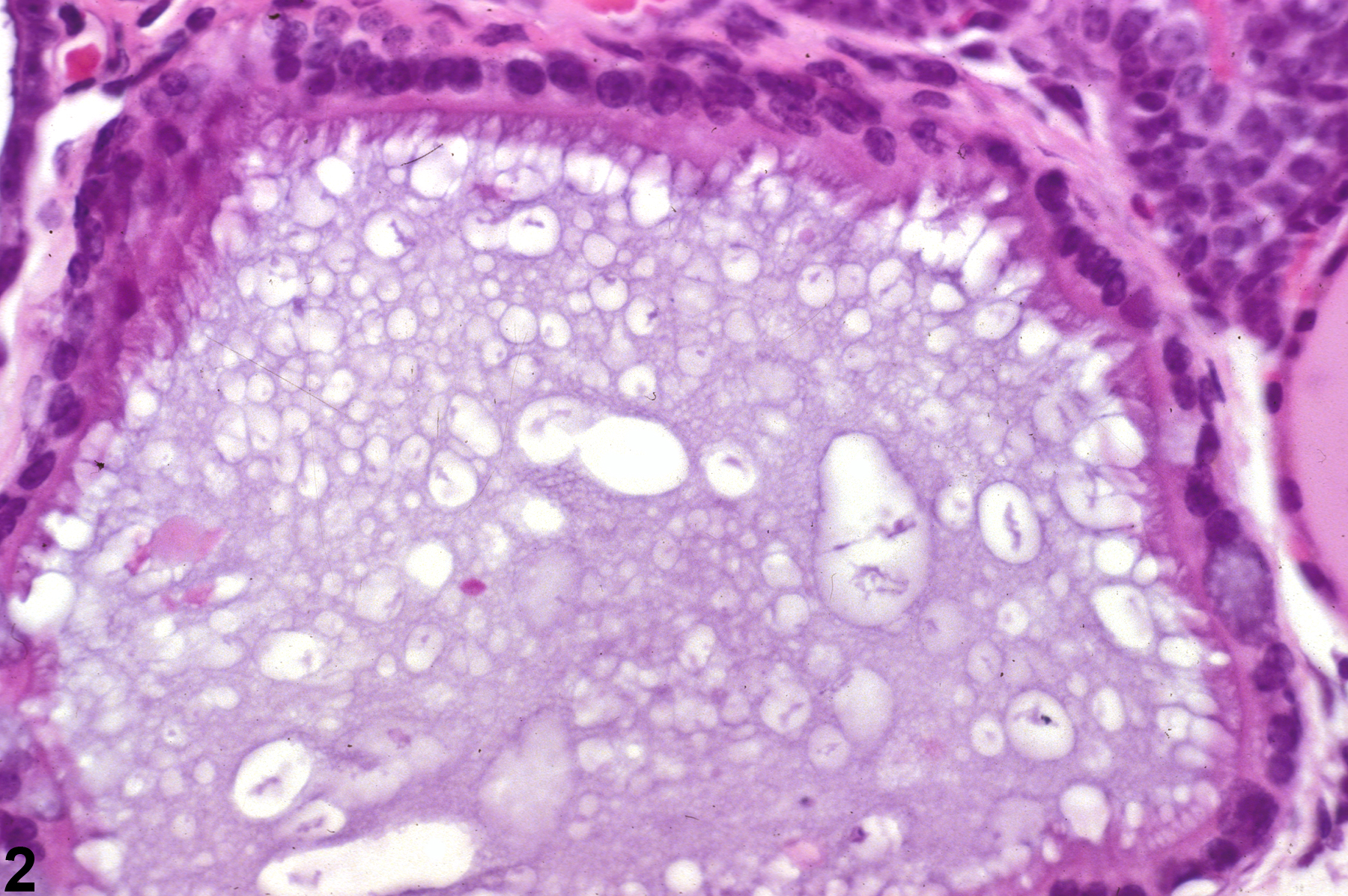
    Parathyroid Gland - Cyst, Congenital in a male B6C3F1 mouse from a chronic study. This higher magnification of Figure 1 shows a cyst lined by ciliated tall cuboidal cells and the presence of vacuolated proteinaceous material filling the cyst lumen.
    Parathyroid Gland - Cyst, Congenital in a male B6C3F1 mouse from a chronic study. This higher magnification of Figure 1 shows a cyst lined by ciliated tall cuboidal cells and the presence of vacuolated proteinaceous material filling the cyst lumen.
    Parathyroid Gland - Cyst, Congenital in a male B6C3F1 mouse from a chronic study. An elongated cyst containing proteinaceous material is present within and extending from the parathyroid.
    Parathyroid Gland - Cyst, Congenital in a male B6C3F1 mouse from a chronic study. An elongated cyst containing proteinaceous material is present within and extending from the parathyroid.
    Parathyroid Gland - Cyst, Congenital in a male B6C3F1 mouse from a chronic study. Higher magnification of Figure 3 showing ciliated columnar cells lining the cyst and flocculant, eosinophilic, proteinaceous material and a degenerating sloughed cell with a nuclear fragment (arrow) in the lumen.
    Parathyroid Gland - Cyst, Congenital in a male B6C3F1 mouse from a chronic study. Higher magnification of Figure 3 showing ciliated columnar cells lining the cyst and flocculant, eosinophilic, proteinaceous material and a degenerating sloughed cell with a nuclear fragment (arrow) in the lumen.

    Authors

    Robert R. Maronpot, DVM, MS, MPH, DACVP, DABT, FIATP
    Senior Pathologist
    Experimental Pathology Laboratories, Inc.
    Research Triangle Park, NC

    Georgette D. Hill, D.V.M., Ph.D.
    Toxicologic Pathologist/Assistant Pathology Program Manager
    Comparative Molecular Pathology Division
    Integrated Laboratory Systems, Inc.
    Research Triangle Park, NC

    Reviewers

    Thomas J. Rosol, DVM, PhD, DACVP
    Professor of Veterinary Biosciences
    Senior Advisor, Life Sciences, Technology Commercialization and Knowledge Transfer
    The Ohio State University
    Columbus, OH

    Amy Brix, DVM, PhD, DACVP
    Senior Pathologist
    Experimental Pathology Laboratories, Inc.
    Research Triangle Park, NC